
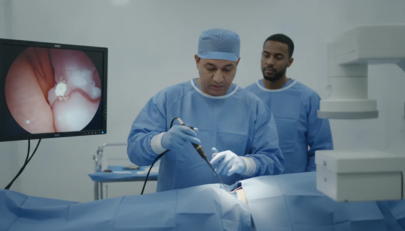
Ureteroscopy
Endoscopic exploration of the ureter and kidney through the natural passages, for diagnostic or therapeutic purposes.
What is it?
When is it indicated?
The ureteroscope travels up through the natural passages to the stone, which is then fragmented with the laser under direct vision, without any incision.
When a CT scan or ultrasound shows an unusual image in the ureter or kidney, endoscopic exploration allows it to be seen directly and its nature clarified.
The presence of blood in the urine must always be explained. Ureteroscopy allows the entire urinary tract to be inspected to find its origin.
If a lesion is discovered on the wall of the ureter or kidney cavities, a small sample is taken at the same time for microscopic analysis.
A narrowed passage in the ureter can obstruct the flow of urine. Some narrowings can be treated directly endoscopically, without open surgery.
Preparation
Procedure
General anesthesia in most cases
Introduction of the ureteroscope through the urethra and then the bladder, without any incision
Advancement under visual control to the area to be explored
Diagnostic or therapeutic procedure as appropriate (laser, biopsy, extraction)
Possible placement of a temporary internal stent
Monitoring in the recovery room
Duration
30 minutes to 1 hour
Aftercare & Recovery
Recovery is generally straightforward, with a return home the same day or the next day. Mild urinary burning or slight bleeding may occur for a few days. The results of any samples taken are communicated at the follow-up consultation.
Risks and side effects
These risks remain rare. Your surgeon will inform you in detail before the procedure.
Request a Quote
Contact us to schedule a consultation or receive a personalized quote.
Request a Quote +216 36 402 000