
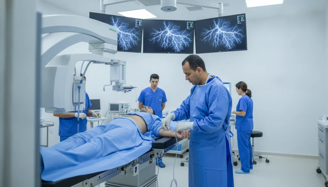
Diagnostic coronary angiography
Centres/Interventional Cardiology/Coronary angiography

Diagnostic coronary angiography

The reference invasive examination for visualizing the coronary arteries by injecting a contrast agent, used to locate and quantify coronary narrowings (stenoses).

What is it?

Coronary angiography is the reference examination ("gold standard") for exploring the coronary arteries, the vessels that supply the heart muscle. It involves inserting a catheter (a thin, flexible tube) into an artery, usually the radial artery at the wrist or the femoral artery at the groin, and guiding it to the openings of the coronary arteries under X-ray guidance. An iodinated contrast agent is then injected to visualize the inside of the coronary arteries on a fluoroscopy screen. This examination provides a precise picture of the anatomy of the coronary arteries and detects and quantifies stenoses (narrowings), occlusions, congenital abnormalities and atheromatous lesions. Coronary angiography is essential for deciding on myocardial revascularization, whether percutaneous (angioplasty with stenting) or surgical (coronary bypass). Coronary angiography is generally performed after non-invasive tests (exercise ECG, stress echocardiography, myocardial scintigraphy) have revealed signs suggestive of coronary artery disease. It is also performed as an emergency in acute coronary syndrome (myocardial infarction) to identify and immediately treat the artery responsible. At the Clinique Pasteur in Tunis, coronary angiography is performed in a cardiac catheterization laboratory equipped with the latest technology. The clinic's interventional cardiologists are highly experienced and perform a large volume of procedures, guaranteeing a high level of quality and safety.
Diagnostic coronary angiography

When is it indicated?

Acute coronary syndrome (heart attack, unstable angina)

In the event of a heart attack or an impending heart attack, this emergency examination immediately identifies the blocked artery so it can be reopened as quickly as possible.

Stable angina resistant to medical treatment

When exertional chest pain persists despite medication, coronary angiography precisely visualizes the narrowings to plan appropriate treatment.

Strongly positive non-invasive tests for myocardial ischemia

When the stress test or stress echocardiography strongly suggests a shortage of blood to the heart, this examination confirms the diagnosis and pinpoints the exact location of the lesions.

Suspected heart failure of ischemic origin

If a weakened heart could be explained by diseased coronary arteries, the examination checks this, because reopening these arteries can improve the heart's strength.

Preoperative assessment before valve surgery in patients at coronary risk

Before operating on a heart valve, the state of the coronary arteries is checked: if they are diseased, they can be treated during the same operation.

Resuscitated cardiac arrest of undetermined origin

After a cardiac arrest of unknown cause, coronary angiography looks for a blocked artery, the most common cause of this type of event.

Assessment of atypical chest pain with multiple risk factors

When unusual pain occurs in a person at risk, this examination provides a definitive answer about the state of the heart's arteries.

Preparation

1Fast strictly for at least 6 hours
2Have a prior blood test (creatinine, coagulation, blood count)
3Report any allergy, particularly to iodinated contrast agents
4Report any anticoagulant or antiplatelet treatment
5Preventive treatment may be prescribed in case of iodine allergy
6Report any history of kidney failure
7Take your usual medications with a little water unless the cardiologist advises otherwise
8Plan for day hospitalization or a short hospital stay (24 hours)
9Arrange for someone to accompany you home

Procedure

1

Positioning on the cardiac catheterization table

2

Disinfection and local anesthesia of the puncture site (wrist or groin)

3

Puncture of the artery and placement of an introducer sheath

4

Insertion of the catheter and advancement to the aorta under X-ray guidance

5

Selective catheterization of the left then the right coronary artery

6

Injection of contrast agent and recording of images from several angles

7

Real-time analysis of the images by the interventional cardiologist

8

Removal of the catheter and the introducer sheath

9

Compression of the puncture site and application of a pressure dressing

10

Post-procedure monitoring with bed rest for 2 to 6 hours depending on the access route

Duration

20 to 45 minutes for the procedure; allow half a day to a full day of hospitalization

Results

Results are communicated immediately after the examination by the interventional cardiologist. The images and the report are provided to the patient and the prescribing physician. A discussion of the treatment strategy takes place if significant lesions are detected.

Risks and side effects

Hematoma at the puncture site (common but usually harmless)
Allergic reaction to the iodinated contrast agent (rare, prevented by premedication)
Coronary dissection or spasm (rare)
Temporary heart rhythm disturbance
Acute kidney failure due to the contrast agent (rare, prevented by hydration)
Stroke (exceptional, less than 0.1%)
Myocardial infarction (exceptional)
Vascular complication at the puncture site (false aneurysm, arteriovenous fistula)

These risks remain rare. Your physician will inform you in detail before the examination.

Book an Appointment

Contact us to schedule your examination or request a quote.

Request a Quote +216 36 402 000

At a glance

Duration: 20 to 45 minutes for the procedure; allow half a day to a full day of hospitalization
Preparation: Required
Book an appointment