
Percutaneous valvuloplasty

Percutaneous valvuloplasty

Percutaneous dilation of a narrowed (stenotic) heart valve using a balloon inflated across the valve, avoiding the need for open-heart surgery.

What is it?

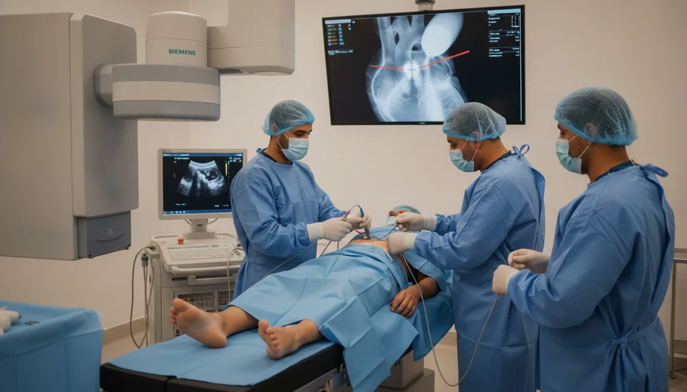
Percutaneous valvuloplasty is an interventional cardiology procedure that dilates a narrowed (stenotic) heart valve using a balloon-tipped catheter. This technique makes it possible to treat certain valve stenoses without resorting to open-heart surgery, offering a less invasive alternative with a faster recovery. Percutaneous mitral valvuloplasty (percutaneous mitral commissurotomy) is the most commonly performed procedure. It is indicated in rheumatic mitral stenosis, a condition still common in Tunisia and the Maghreb countries. The technique involves inserting a special balloon (Inoue balloon) through the femoral vein, crossing the interatrial septum by transseptal puncture, then inflating the balloon at the mitral valve to separate the fused commissures. Percutaneous aortic valvuloplasty can also be performed in severe aortic stenosis, mainly as palliative treatment or as a bridge to aortic valve replacement (surgical or transcatheter - TAVI). The technique involves inserting a balloon retrogradely through the femoral artery and inflating it at the aortic valve. At the Clinique Pasteur in Tunis, percutaneous valvuloplasties are performed by interventional cardiologists with recognized expertise in percutaneous valve procedures. The clinic has a complete technical platform, including intraprocedural transesophageal echocardiography to guide and optimize the result.
Percutaneous valvuloplasty

When is it indicated?

Symptomatic rheumatic mitral stenosis with favorable valve anatomy

When the mitral valve, narrowed as a result of rheumatic fever, causes breathlessness and fatigue, it can be widened with a balloon if its shape allows, without opening the chest.

Tight mitral stenosis with a valve area of less than 1.5 cm2

When the valve opening becomes very narrow, blood struggles to flow into the heart; dilation restores an adequate passage through the valve.

Mitral stenosis with pulmonary arterial hypertension

A narrowed mitral valve can raise the pressure in the lungs; widening the valve relieves this pressure and protects the right side of the heart.

Mitral stenosis in pregnant women (alternative to surgery)

During pregnancy, this gentle technique treats a poorly tolerated valve narrowing while avoiding open-heart surgery, which is harder on mother and baby.

Severe symptomatic aortic stenosis in inoperable patients (palliative treatment)

In people too frail for surgery, dilating the aortic valve relieves symptoms and improves quality of life.

Severe aortic stenosis as a bridge to TAVI or surgical replacement

Dilation can serve as an intermediate step: it quickly relieves the patient while awaiting definitive valve replacement under better conditions.

Severe congenital pulmonary stenosis

When the pulmonary valve has been narrowed since birth, the balloon opens it effectively and usually avoids the need for surgery.

Preparation

1Fast strictly for at least 8 hours
2Complete blood tests (full blood count, coagulation, creatinine, blood group, antibody screening)
3Recent transthoracic and transesophageal echocardiograms
4Rule out the presence of a thrombus in the left atrium (TEE mandatory before mitral valvuloplasty)
5Report any allergy, particularly to iodinated contrast agents
6Stop oral anticoagulants according to the cardiologist's instructions
7Hospitalization is required (2 to 4 days)
8A signed informed consent form is mandatory
9Dental check-up to rule out any source of infection
10Cardiac CT scan if indicated

Procedure

1

Positioning in the catheterization room under local anesthesia and light sedation

2

Continuous monitoring (ECG, blood pressure, oxygen saturation)

3

Intraprocedural transesophageal echocardiography for guidance

4

Femoral venous puncture and placement of the introducer sheath

5

For the mitral valve: transseptal puncture to access the left atrium

6

Administration of intravenous heparin

7

Advancement and positioning of the balloon catheter at the stenotic valve

8

Gradual inflation of the balloon to dilate the valve

9

Immediate assessment of the result by echocardiography and measurement of pressure gradients

10

If necessary, further inflation(s) with a slightly larger balloon volume

11

Final hemodynamic and echocardiographic check

12

Removal of the equipment and hemostasis of the puncture site

13

Transfer to the intensive care unit for postoperative monitoring

Duration

1 to 2 hours for the procedure; hospitalization for 2 to 4 days

Results

The result is assessed immediately during the procedure by echocardiography and measurement of pressure gradients. The reduction in the stenosis is usually significant from the first inflation. An echocardiographic check is performed the next day and a follow-up consultation is scheduled at 1 month.

Risks and side effects

Significant mitral or aortic regurgitation (the main complication, which may require surgery)
Cardiac tamponade due to perforation (rare, requiring emergency drainage)
Systemic embolism (stroke or peripheral embolism)
Heart rhythm disturbance (atrial fibrillation, atrioventricular block)
Hematoma or vascular complication at the puncture site
Residual atrial septal defect after transseptal puncture (usually of no consequence)
Allergic reaction to the contrast agent
Kidney failure due to the contrast agent
Valve restenosis in the medium or long term (requiring a repeat procedure)
Periprocedural death (exceptional, less than 0.5%)

These risks remain rare. Your physician will inform you in detail before the examination.

Book an Appointment

Contact us to schedule your examination or request a quote.

Request a Quote +216 36 402 000

At a glance

Duration: 1 to 2 hours for the procedure; hospitalization for 2 to 4 days
Preparation: Required
Book an appointment