
Right and left heart catheterization
Centres/Interventional Cardiology/Cardiac catheterization

Right and left heart catheterization

Invasive hemodynamic exploration of the right and left heart chambers by catheterization, measuring intracardiac pressures and blood flow to assess overall cardiac function.

What is it?

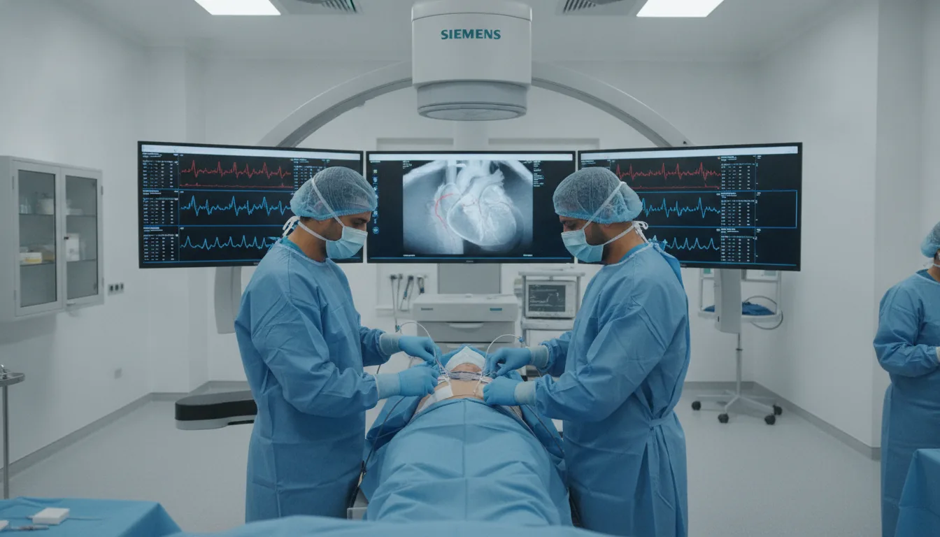
Right and left heart catheterization is an invasive hemodynamic examination that involves inserting catheters into the heart chambers to directly measure intracardiac pressures, blood flow and vascular resistance. Right heart catheterization explores the right atrium, the right ventricle and the pulmonary artery, while left heart catheterization explores the aorta and the left ventricle. Right heart catheterization is performed by inserting a catheter (usually a Swan-Ganz catheter) through a vein, typically the internal jugular vein, the subclavian vein or the femoral vein. This catheter is guided to the pulmonary artery and measures the pressures in the right chambers, the pulmonary capillary wedge pressure (an indirect reflection of the pressure in the left atrium), cardiac output and pulmonary vascular resistance. Left heart catheterization is performed via an arterial (retrograde) route or a transseptal route. It measures the pressures in the aorta and the left ventricle, calculates transvalvular gradients (in cases of aortic or mitral stenosis) and assesses the contractile function of the left ventricle by ventriculography (injection of contrast into the ventricle). At the Clinique Pasteur in Tunis, cardiac catheterization is performed by highly qualified interventional cardiologists in a catheterization laboratory equipped with the most advanced technology. Each procedure is tailored to the patient's specific indication in order to obtain the hemodynamic information needed for treatment decisions.
Right and left heart catheterization

When is it indicated?

Pulmonary arterial hypertension: diagnosis and assessment of severity

When the pressure is too high in the arteries of the lungs, only this direct measurement can confirm the disease and gauge its severity.

Severe heart failure: hemodynamic assessment before transplantation

In a patient whose heart is severely weakened, the examination precisely measures pressures and blood flow to prepare major treatment decisions.

Severe valve disease: assessment of the hemodynamic impact

When a valve is badly damaged, these direct measurements clarify the real impact on heart function and help choose the best time to intervene.

Restrictive cardiomyopathy versus constrictive pericarditis

Two diseases that stiffen the heart can look very similar; studying the pressures inside the heart distinguishes them and guides treatment.

Complex congenital heart disease: assessment of shunts and pressures

In cases of heart malformation present from birth, the examination measures abnormal blood flow between the chambers and guides the correction strategy.

Pre-heart transplant assessment

Before a heart transplant, these highly precise measurements check in particular the state of the pulmonary circulation, an essential condition for the success of the transplantation.

Cardiogenic shock: support for treatment decisions

In situations where the heart can no longer maintain adequate circulation, continuous pressure monitoring helps the team adjust treatments in real time.

Assessment of the response to pulmonary vasodilators

The examination directly tests the effect of medications designed to relax the arteries of the lungs, in order to choose the most effective treatment for each patient.

Preparation

1Fast strictly for at least 6 hours
2Complete prior blood tests (creatinine, coagulation, full blood count)
3Report any allergy, particularly to contrast agents
4Report all current treatments, particularly anticoagulants
5Hospitalization is generally required (24 to 48 hours)
6Bring recent ECG and echocardiography results
7Inform the doctor of any history of kidney failure

Procedure

1

Positioning on the catheterization table under continuous monitoring (ECG, blood pressure, saturation)

2

Disinfection and local anesthesia of the puncture site(s)

3

For right heart catheterization: venous puncture (jugular, subclavian or femoral)

4

Insertion of the Swan-Ganz catheter and advancement through the right chambers

5

Measurement of pressures in the right atrium, the right ventricle and the pulmonary artery

6

Measurement of the pulmonary capillary wedge pressure and cardiac output

7

For left heart catheterization: arterial puncture (radial or femoral)

8

Insertion of the catheter and retrograde advancement to the left ventricle

9

Measurement of aortic and left ventricular pressures

10

Left ventriculography if necessary (injection of contrast)

11

Calculation of vascular resistance, blood flow and hemodynamic indices

12

Removal of the catheters and compression of the puncture sites

Duration

45 minutes to 1h30 depending on the extent of the exploration; hospitalization for 24 to 48 hours

Results

The hemodynamic results are available immediately. A full report with all the pressure, flow and resistance measurements is written and discussed with the medical team to guide the treatment strategy.

Risks and side effects

Hematoma at the puncture site
Heart rhythm disturbance as the catheter passes through (usually temporary)
Pneumothorax in case of subclavian or jugular puncture (rare)
Cardiac perforation (exceptional)
Infection at the puncture site (rare with aseptic precautions)
Vagal reaction (faintness, slowing of the heart)
Air embolism (exceptional)
Reaction to the contrast agent (if ventriculography is performed)

These risks remain rare. Your physician will inform you in detail before the examination.

Book an Appointment

Contact us to schedule your examination or request a quote.

Request a Quote +216 36 402 000

At a glance

Duration: 45 minutes to 1h30 depending on the extent of the exploration; hospitalization for 24 to 48 hours
Preparation: Required
Book an appointment