
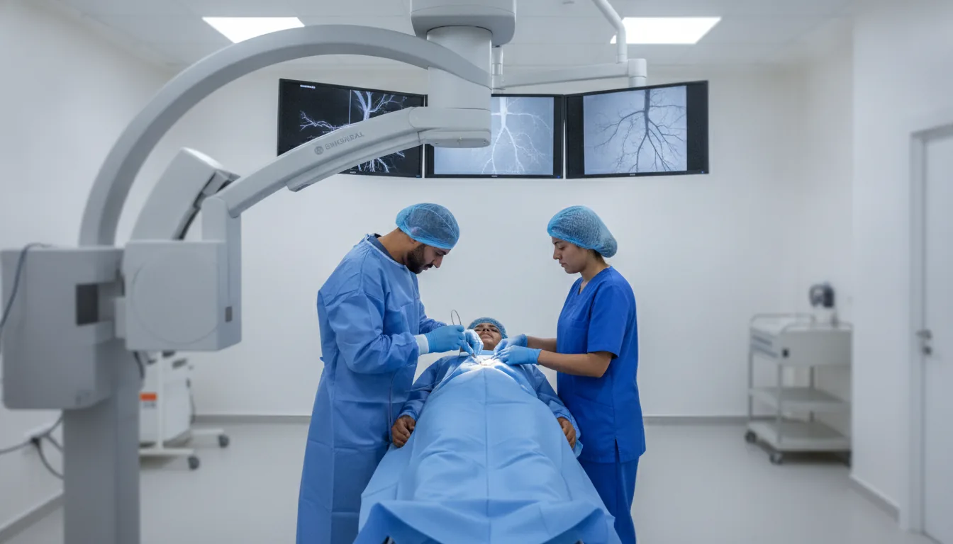
Interventional radiology - embolization, drainage, radiofrequency
Minimally invasive image-guided therapeutic procedures, including embolization, drainage and radiofrequency thermal ablation, offering less demanding alternatives to conventional surgery.
What is it?
When is it indicated?
When fibroids cause heavy bleeding or pain, this technique shrinks them by blocking their blood supply, without an operation and while preserving the uterus.
In cases of severe bleeding, the radiologist can block the bleeding vessel from the inside using a fine catheter. This rapid procedure often stops the hemorrhage without open surgery.
This treatment delivers a medication directly into the liver tumor while blocking the vessels that feed it, concentrating the effect on the lesion while sparing the rest of the organ.
A fine needle placed at the heart of the tumor under imaging guidance destroys it with heat. This option is invaluable for patients for whom surgery is not advisable.
Rather than operating, the radiologist can evacuate a pocket of infection by placing a small drain through the skin, guided by imaging. The patient is thus relieved with a minimally invasive procedure.
Some blood vessel abnormalities carry a risk of bleeding. Embolization makes it possible to close them from the inside, through the natural pathways of the bloodstream, and eliminate this risk.
The injection of medical cement into a fractured, painful vertebra consolidates it quickly. This procedure relieves the pain and allows mobility to be regained more rapidly.
When bile or urine can no longer flow normally, a drain placed through the skin restores their drainage, relieves the patient and protects the organ concerned.
Preparation
Procedure
The patient is positioned in the intervention room under cardiac and blood pressure monitoring
A peripheral IV line is placed for the administration of medications and sedatives
Local anesthesia is performed at the puncture site (general anesthesia possible depending on the procedure)
For embolization: a catheter is introduced through the femoral artery and guided under fluoroscopy to the target vessel
For drainage: a needle and then a drain are placed under ultrasound or CT guidance into the fluid collection
For radiofrequency: a needle electrode is positioned in the tumor under CT guidance
The procedure is performed under continuous imaging control
A final check is carried out to verify the effectiveness of the treatment
The patient is monitored in the recovery room and then during hospitalization
Duration
45 minutes to 2 hours depending on the complexity of the procedure
Results
The interventional radiologist informs the patient and the referring physician of how the procedure went immediately afterwards. A detailed report is written. A follow-up imaging examination is scheduled in the following weeks to assess the effectiveness of the treatment.
Risks and side effects
These risks remain rare. Your physician will inform you in detail before the examination.
Book an Appointment
Contact us to schedule your examination or request a quote.
Request a Quote +216 36 402 000