
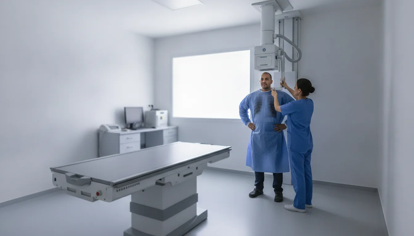
Bone radiology - flat-panel bone and chest tables
Centres/Radiology/Bone radiology

Bone radiology - flat-panel bone and chest tables

High-resolution digital radiography using flat-panel detector technology for exploration of the skeleton and lungs, offering superior image quality with a minimal radiation dose.

What is it?

Bone radiology on a table equipped with flat-panel detectors represents the most recent evolution of conventional radiography. The digital flat-panel detector replaces the old film cassettes or phosphor plates with an electronic detector built into the table, capable of directly converting X-rays into high-resolution digital images. This technology offers markedly superior image quality while significantly reducing the radiation dose. The flat-panel detector allows instant image acquisition without processing delay, a wider exposure dynamic range (fewer retakes), advanced image post-processing (contrast adjustment, zoom, measurements) and direct integration into the digital archiving system (PACS). These advantages translate into time savings for the patient and optimal diagnostic quality for the radiologist. This technique is used as a first-line examination for exploring the entire skeleton (fractures, osteoarthritis, bone tumors, malformations), the lungs and chest (pneumonia, pneumothorax, pleural effusions, tumors), as well as the abdomen (intestinal obstruction, calcifications, foreign bodies). Radiography remains the most frequently prescribed imaging examination in the world and is often the first step in the diagnostic workup. At Clinique Pasteur in Tunis, the radiology rooms are equipped with latest-generation flat-panel tables, guaranteeing optimal image quality for a reliable diagnosis. The digital system allows rapid image transmission and secure archiving in the computerized patient record.
Bone radiology - flat-panel bone and chest tables

When is it indicated?

Fractures and bone trauma

After a fall or an impact, an X-ray is the first examination performed: it immediately shows whether a bone is broken, cracked or displaced, and guides the next steps of care.

Osteoarthritis and degenerative joint diseases

When a joint becomes painful or stiff over time, an X-ray makes it possible to assess cartilage wear and changes in the bones, in order to adapt the treatment.

Primary bone tumors and metastases

In the presence of unusual bone pain or as part of a cancer workup, an X-ray looks for abnormal areas in the bones and points to further examinations if needed.

Bone infections (osteomyelitis, septic arthritis)

When an infection of the bone or a joint is suspected, for example in the presence of pain associated with fever, an X-ray helps identify its signs and monitor its progression under treatment.

Bone malformations and growth disorders

In children as in adults, an X-ray makes it possible to study the shape and alignment of the bones, for example to monitor scoliosis or check that growth is progressing normally.

Lung conditions: pneumonia, pneumothorax, tumor, pleural effusion

A chest X-ray is the basic examination for a lingering cough, fever, shortness of breath or chest pain: it quickly provides an overall view of the lungs and heart.

Preoperative radiological assessment

Before certain operations, the surgeon requests X-rays to plan the procedure precisely, for example to choose and position a joint prosthesis.

Postoperative monitoring of fixation hardware or prostheses

After the placement of screws, plates or a prosthesis, regular X-rays check that the hardware remains properly in place and that the bone is healing correctly.

Preparation

1No special preparation in most cases
2Remove clothing and metallic objects from the area to be X-rayed (jewelry, belt, piercings)
3Report any pregnancy or suspected pregnancy
4Bring the doctor's prescription specifying the region to be explored
5Bring previous X-rays for comparison if available
6For abdominal X-rays, fasting may be recommended
7Wear clothing that is easy to remove

Procedure

1

The radiology technologist positions the patient according to the region to be explored (standing, lying down or seated)

2

A lead apron protects sensitive areas not being examined (gonads, thyroid)

3

The patient must remain still while the image is taken (a few seconds)

4

Breathing instructions are given: breath held for chest X-rays

5

The image appears instantly on the control screen thanks to the flat-panel detector

6

Several views may be necessary (frontal, lateral, oblique)

7

The technologist checks the quality of the images before releasing the patient

8

The images are sent to the radiologist for interpretation

Duration

5 to 15 minutes depending on the number of images required

Results

The digital images are available immediately. The radiologist's report is generally available within 24 hours. In emergencies, the results are communicated immediately to the treating physician.

Risks and side effects

Exposure to X-rays (very low dose, optimized by the flat-panel detector)
No pain or side effects
Relative contraindication in case of pregnancy (lead shielding of the fetus if the examination is essential)
Theoretical risk related to ionizing radiation negligible for a single examination

These risks remain rare. Your physician will inform you in detail before the examination.

Book an Appointment

Contact us to schedule your examination or request a quote.

Request a Quote +216 36 402 000

At a glance

Duration: 5 to 15 minutes depending on the number of images required
Preparation: Required
Book an appointment