
Mammography
Specialized radiological examination of the breast allowing early breast cancer screening and the diagnosis of breast abnormalities, using a controlled compression technique for high-resolution images.
What is it?
When is it indicated?
Even without any symptoms, regular mammography can detect possible abnormalities at a very early stage, when they are still invisible and impalpable, which considerably increases the chances of recovery.
If you or your doctor have noticed a lump, a discharge or a change in the skin of the breast, mammography helps determine its nature and decide on any additional examinations.
After treatment, regular mammograms check that the tissues are healing properly and allow any new abnormality to be detected very early, for reassuring follow-up.
Before a cosmetic breast procedure, a mammogram makes sure there is no underlying abnormality, so that the surgery can take place under the safest possible conditions.
Breast pain that persists is most often benign, but mammography makes it possible to look for its cause and reassure the patient by ruling out an abnormality of the breast tissue.
Some findings considered very probably benign are simply monitored closely: regular check-ups verify that they remain stable over time.
When close relatives have been affected by breast cancer, earlier and more frequent monitoring is offered in order to detect the slightest abnormality as early as possible.
Preparation
Procedure
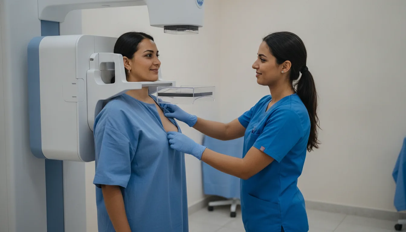
The technologist positions the breast on the mammography unit plate
The breast is compressed between two plates gradually and in a controlled manner
Two standard views are taken of each breast: frontal (craniocaudal) and oblique (mediolateral oblique)
The compression lasts a few seconds while the image is acquired
Additional views may be taken if needed (magnification, localized compression)
The radiologist checks the quality of the images and may request additional views
The entire examination is generally completed in less than 15 minutes
Duration
10 to 15 minutes
Results
The images are analyzed by a radiologist specialized in breast imaging. The results are classified according to the BI-RADS classification (from 0 to 6) and are generally available within 24 to 48 hours. If an abnormality is found, additional examinations are offered promptly.
Risks and side effects
These risks remain rare. Your physician will inform you in detail before the examination.
Book an Appointment
Contact us to schedule your examination or request a quote.
Request a Quote +216 36 402 000