
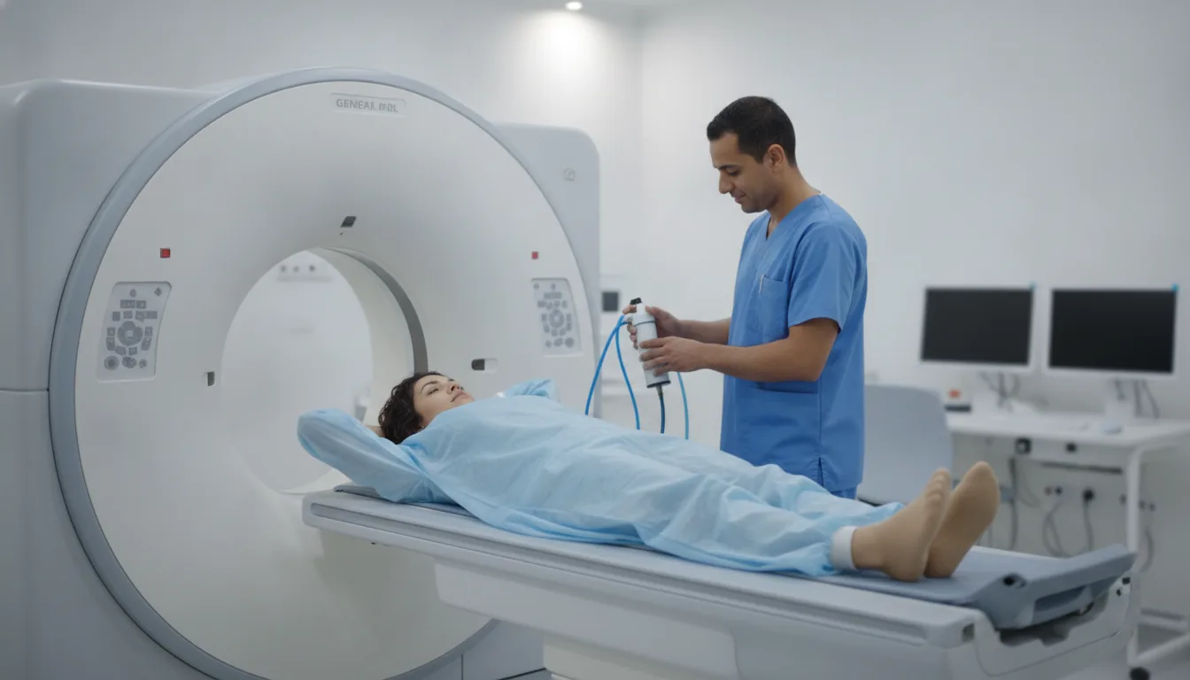
Dual-energy CT scan

Dual-energy CT scan

Latest-generation CT scanner using dual-energy technology for superior tissue differentiation, enabling more accurate diagnoses with an optimized radiation dose.

What is it?

The dual-energy CT scanner, also known as spectral computed tomography, represents a major advance in the field of medical imaging. This innovative technology simultaneously uses two different X-ray energy levels, allowing spectral analysis of the tissues being imaged. This approach offers diagnostic capabilities considerably superior to those of conventional single-energy CT. The principle of dual-energy CT is based on the fact that the different tissues and materials in the human body absorb X-rays differently depending on their energy level. By combining the information obtained at two distinct energies, the scanner can differentiate structures that appear identical on a conventional CT scan: for example, distinguishing a urate stone from a calcium oxalate stone, identifying the composition of an atheromatous plaque, or differentiating a recent hematoma from an old one. This technology also makes it possible to significantly reduce metal artifacts related to prostheses, to improve the visualization of tumor contrast enhancement, to create virtual maps without injection and to reduce the radiation dose delivered to the patient. Dual-energy CT is particularly effective in oncology, urology, cardiology and vascular disease. At Clinique Pasteur in Tunis, dual-energy CT is performed on a latest-generation machine offering exceptional spatial and temporal resolution. The team of expert radiologists makes full use of the capabilities of this technology to provide accurate and rapid diagnoses, with constant attention to patient radiation protection.
Dual-energy CT scan

When is it indicated?

Cancer assessment and follow-up: detection, staging, evaluation of treatment response

The CT scan makes it possible to locate a lesion, measure its extent and monitor its progression during treatment. It is an essential tool for the medical team to adapt care at every stage.

Vascular conditions: CT angiography, pulmonary embolism, aortic dissection

With a contrast agent injection, the CT scan visualizes the blood vessels in detail. It is requested urgently when a clot in the lungs or damage to the aorta is suspected.

Chest conditions: lung nodules, pneumonia, mediastinum

In cases of persistent cough, shortness of breath or an abnormality on an X-ray, the chest CT scan finely examines the lungs and the region between them to determine the cause.

Abdominal and pelvic conditions: liver, pancreas, kidneys, colon

The CT scan explores all the organs of the abdomen in a matter of moments. It helps explain abdominal pain, characterize an abnormality seen on ultrasound or look for an infection.

Urological conditions: urinary stones with composition analysis

The CT scan detects stones in the kidneys and urinary tract with great reliability. Dual-energy technology can even help determine their nature, which guides treatment to prevent recurrence.

Bone and joint conditions: complex fractures, detection of bone metastases

When a fracture is difficult to analyze on a plain X-ray, the CT scan provides three-dimensional images of the bones. It is also used to check the condition of the skeleton as part of a cancer workup.

Cardiac assessment: coronary CT angiography, calcium score

Cardiac CT makes it possible to examine the arteries of the heart without any intervention and to assess the presence of calcium deposits, helping the cardiologist estimate the health of the coronary arteries.

Emergencies: trauma, stroke, acute abdomen

Thanks to its speed, the CT scan is the examination of choice in emergency situations: it quickly assesses the situation after an accident, in the presence of signs of stroke or sudden abdominal pain.

Preparation

1Report any known allergy, particularly to iodinated contrast agents
2Report any kidney failure or known kidney disease
3Fast for 4 hours if a contrast agent injection is planned
4A recent kidney function test (creatinine, clearance) is required before injection
5Report any pregnancy or suspected pregnancy (absolute contraindication)
6Report current treatments, particularly metformin (diabetes)
7Remove metallic objects from the area being examined
8Bring the medical prescription and previous examinations

Procedure

1

The patient is positioned on the scanner table, generally lying on their back

2

A scout view (localization image) is taken first

3

If an injection is needed, a catheter is placed in a vein of the arm

4

The iodinated contrast agent is injected intravenously using an automatic injector

5

The table moves through the scanner ring during acquisition

6

Several passes may be necessary (before and after injection, at different phases)

7

The patient must remain still and follow the breathing instructions (breath held)

8

The examination is painless; only a sensation of warmth is felt during the injection

Duration

10 to 20 minutes (rapid acquisition, a few seconds per pass)

Results

The images are reconstructed and analyzed by the radiologist. The report is generally available within 24 to 48 hours. In emergencies, the results are communicated immediately to the referring physician.

Risks and side effects

Exposure to X-rays (dose optimized thanks to dual-energy technology)
Allergic reaction to the iodinated contrast agent (hives, edema, anaphylactic shock - rare)
Acute kidney failure related to the contrast agent (contrast-induced nephropathy - rare)
Extravasation of the contrast agent at the injection site (local pain, generally benign)
Absolute contraindication in case of pregnancy
Interaction with metformin (temporary discontinuation for 48h after injection in diabetic patients)

These risks remain rare. Your physician will inform you in detail before the examination.

Book an Appointment

Contact us to schedule your examination or request a quote.

Request a Quote +216 36 402 000

At a glance

Duration: 10 to 20 minutes (rapid acquisition, a few seconds per pass)
Preparation: Required
Book an appointment