
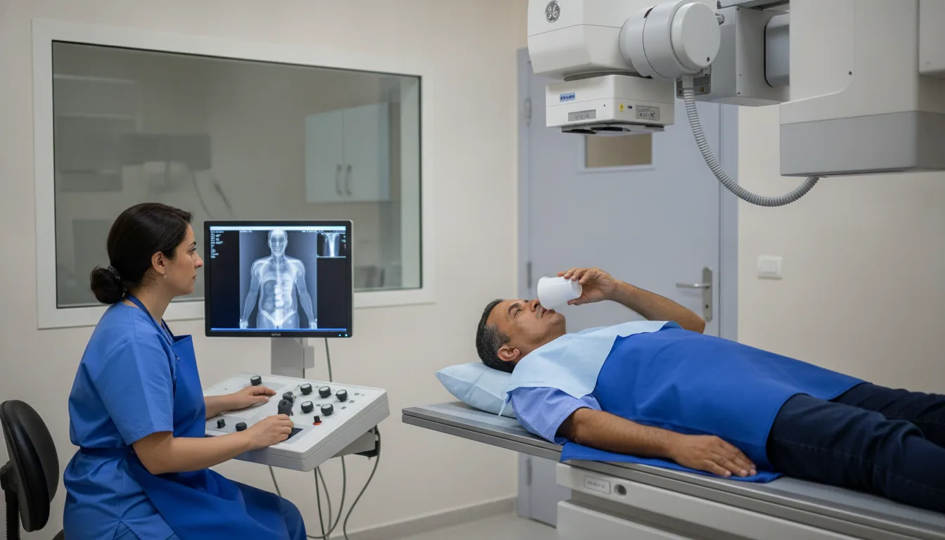
Conventional radiology - remote-controlled table
Centres/Radiology/Conventional radiology

Conventional radiology - remote-controlled table

Radiological examination performed on a remote-controlled table allowing real-time dynamic explorations, particularly digestive transit studies and examinations with contrast opacification.

What is it?

Conventional radiology on a remote-controlled table is an imaging technique that combines the principles of classic radiography with the ability to perform dynamic examinations in real time. The remote-controlled table allows the radiologist to change the patient's position and the angle of the X-ray beam from a distance, while viewing the images live on a screen, thus providing a complete and precise exploration of the anatomical structures. This technique is particularly suited to explorations of the digestive tract (upper gastrointestinal series, barium enema, small bowel follow-through), urological examinations (intravenous urography, cystography), arthrography and hysterosalpingography. The use of contrast agents (barium or iodine) makes it possible to visualize in real time the passage of the agent through the hollow organs, revealing any morphological or functional abnormalities. The modern remote-controlled table is equipped with a digital fluoroscopy system that offers excellent image quality while significantly reducing the radiation dose delivered to the patient. The radiologist controls the entire examination from the control console, guiding the movements of the table and the image acquisitions according to real-time findings. At Clinique Pasteur in Tunis, the latest-generation remote-controlled table makes it possible to perform the full range of conventional radiology examinations with contrast under optimal conditions of quality and safety. The team of experienced radiologists adapts the protocol to each clinical situation for an accurate diagnosis.
Conventional radiology - remote-controlled table

When is it indicated?

Upper gastrointestinal series: reflux, ulcer, hiatal hernia, tumor

After drinking a contrast agent, the radiologist follows its path through the esophagus and stomach live. This examination helps explain heartburn, acid reflux or difficulty swallowing.

Small bowel follow-through: Crohn's disease, obstruction, tumor

This examination follows the progression of the contrast agent through the small intestine, an area that is difficult to access. It is useful for investigating pain, chronic inflammation or slowed transit.

Barium enema: polyps, colon tumor, diverticulosis

The contrast agent introduced rectally outlines the contours of the large intestine on the images. It makes it possible to detect irregularities of the colon wall when colonoscopy is not feasible.

Cystography: vesicoureteral reflux, incontinence, bladder tumor

By filling the bladder with contrast agent, the radiologist observes how it functions during filling and emptying. This examination helps explain recurrent urinary infections or urinary leakage.

Hysterosalpingography: infertility workup, tubal patency

This examination checks that the fallopian tubes are not blocked and that the uterine cavity has a normal shape. It is part of the workup offered to couples who are having difficulty conceiving.

Arthrography: joint injuries before MRI or as a complement

A contrast agent injected into the joint makes fine structures such as cartilage or ligaments visible. This examination sometimes complements MRI to better analyze a painful joint.

Exploration of swallowing and digestive fistulas

By filming the passage of the contrast agent in real time, the radiologist analyzes how the patient swallows and looks for any abnormal passages in the digestive tract, for example after surgery.

Preparation

1Fast for 6 hours before digestive explorations
2For barium enema: bowel preparation according to the instructions provided (low-residue diet, laxatives)
3Report any allergy to contrast agents (barium, iodine)
4Report any pregnancy or suspected pregnancy
5Report any history of digestive surgery
6Bring the doctor's prescription and previous examinations
7Wear clothing that is easy to remove and free of metallic elements

Procedure

1

The patient is positioned on the remote-controlled table

2

The radiologist explains how the examination will proceed and the instructions to follow

3

A contrast agent is administered orally, rectally or intravenously depending on the examination

4

The radiologist follows the progression of the contrast agent in real time on the screen (fluoroscopy)

5

The table is tilted into different positions to visualize all the structures

6

X-ray images are taken at key moments of the examination

7

The patient must follow the radiologist's positioning and breathing instructions

8

At the end of the examination, additional images may be necessary

Duration

15 to 45 minutes depending on the type of exploration

Results

The images are analyzed by the radiologist, who writes a report. Results are available within 24 to 48 hours. In case of emergency, the results are communicated immediately.

Risks and side effects

Exposure to X-rays (reduced dose thanks to digital fluoroscopy)
Transient constipation after barium ingestion (drink plenty of fluids after the examination)
Allergic reaction to the iodinated contrast agent (rare)
Digestive perforation during a barium enema (exceptional)
Absolute contraindication in case of pregnancy
Aspiration of the contrast agent in case of swallowing disorders

These risks remain rare. Your physician will inform you in detail before the examination.

Book an Appointment

Contact us to schedule your examination or request a quote.

Request a Quote +216 36 402 000

At a glance

Duration: 15 to 45 minutes depending on the type of exploration
Preparation: Required
Book an appointment