
MRI - Magnetic Resonance Imaging
Medical imaging technique using a powerful magnetic field and radiofrequency waves to obtain detailed images of the body's organs and tissues, without ionizing radiation.
What is it?
When is it indicated?
MRI is the most accurate examination for observing the brain. It helps the doctor understand the cause of unusual headaches, memory problems, dizziness or seizures, and check the condition of brain tissue.
In cases of persistent back or neck pain, or pain radiating down an arm or a leg, MRI shows the discs, nerves and spinal cord to identify what is compressing or irritating these structures.
After a sprain, a sports injury or long-lasting joint pain, MRI finely visualizes the menisci, ligaments and cartilage of the knee, shoulder or other joints, where X-rays only show the bones.
When a cancer is diagnosed, MRI helps determine the size of the lesion and check whether other areas are affected. It is also used to monitor the effectiveness of treatments over time.
MRI allows detailed study of the liver and bile ducts, for example to characterize an abnormality found on ultrasound or to look for a stone in the bile ducts, without any radiation.
For the organs of the pelvis, MRI provides very precise images. It is requested to investigate pelvic pain, a fibroid, a cyst, or for an assessment of the prostate or rectum.
This technique visualizes the arteries and veins without any intervention. It is useful for checking proper blood circulation and looking for a narrowing or dilation of a vessel.
Cardiac MRI studies the heart muscle in detail: it assesses its contraction, its blood supply and the condition of the tissue after a cardiac event, helping the cardiologist choose the most appropriate treatment.
Preparation
Procedure
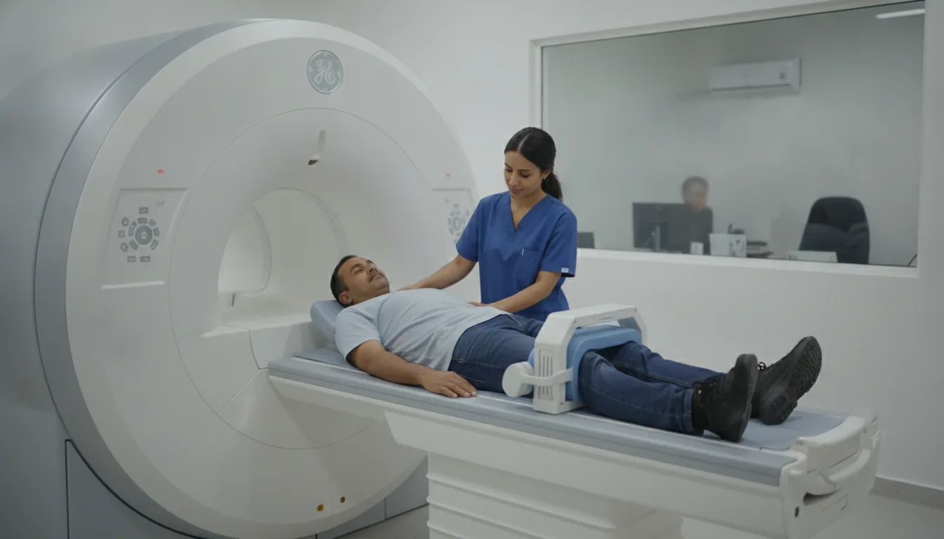
The patient is positioned on the examination table, lying on their back in most cases
A coil is placed over the region to be examined to improve image quality
The table slides inside the magnet (tunnel)
The patient must remain still throughout the entire examination
Loud, repetitive noises are emitted by the machine (earplugs or headphones are provided)
An intercom allows communication with the medical team at any time
If a contrast agent (gadolinium) is injected, an IV line is placed in the arm
Several acquisition sequences are performed, interspersed with short pauses
Duration
20 to 45 minutes depending on the region explored and the protocol
Results
The images are analyzed by the radiologist, who writes a detailed report. Results are generally available within 24 to 48 hours and sent to the referring physician.
Risks and side effects
These risks remain rare. Your physician will inform you in detail before the examination.
Book an Appointment
Contact us to schedule your examination or request a quote.
Request a Quote +216 36 402 000