
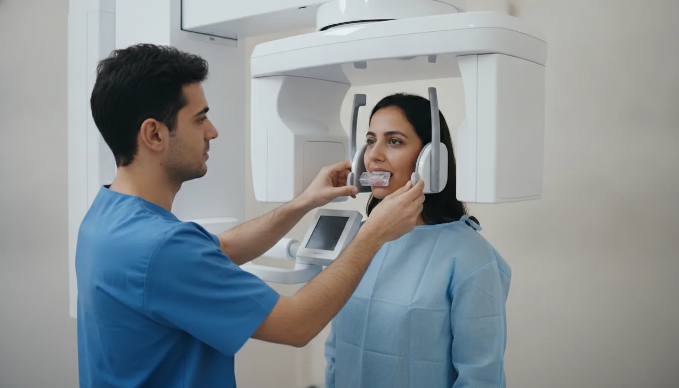
2D dental panoramic X-ray
Panoramic X-ray of the upper and lower dental arches in a single image, providing an overall view of the teeth, jaws and adjacent structures.
What is it?
When is it indicated?
In a single image, the dentist visualizes all the teeth and the bone that supports them. This makes it possible to detect hidden cavities, infections at the root of the teeth or early gum recession.
Before removing wisdom teeth, the panoramic X-ray shows their position, the orientation of their roots and their proximity to the nerves, allowing the practitioner to plan a safe and precise procedure.
Before implant placement or orthodontic treatment, this overall view makes it possible to assess the amount of bone available and the position of all the teeth in order to properly prepare the treatment.
Some teeth remain trapped in the bone or develop in excess without being visible in the mouth. The panoramic X-ray locates them precisely, particularly in children and adolescents.
After an impact to the face, this quick examination checks the integrity of the jaw and teeth, and makes it possible to detect a possible fracture.
The panoramic X-ray can reveal lesions developing in the jawbone, which are often silent, and point to more detailed imaging if necessary.
In cases of jaw pain, clicking or difficulty opening the mouth, this examination gives a first overview of the joints connecting the jaw to the skull.
Some sinus infections originate from an infected tooth whose root is close to the sinus. The panoramic X-ray helps identify the tooth responsible so the problem can be treated at its source.
Preparation
Procedure
The patient stands facing the machine
They bite on a positioning rod that holds the dental arches in the correct position
The forehead and temples are held by supports to prevent any movement
The arm of the machine rotates around the patient's head (about 15 seconds)
The patient must remain perfectly still, with the tongue pressed against the palate and the lips closed
The panoramic image appears immediately on the screen in digital form
The radiologist checks the quality of the image and interprets it
Duration
5 minutes (acquisition takes a few seconds)
Results
The digital image is available immediately. The radiology report is sent to the referring dentist or surgeon within 24 hours. The patient can obtain a digital copy of their panoramic X-ray.
Risks and side effects
These risks remain rare. Your physician will inform you in detail before the examination.
Book an Appointment
Contact us to schedule your examination or request a quote.
Request a Quote +216 36 402 000