
Bone densitometry
Centres/Radiology/Bone densitometry

Bone densitometry

Measurement of bone mineral density by dual-energy X-ray absorptiometry (DEXA), the reference examination for the diagnosis of osteoporosis and the assessment of fracture risk.

What is it?

Bone densitometry, also called dual-energy X-ray absorptiometry (DEXA), is the reference examination for measuring bone mineral density (BMD). This precise and reproducible technique makes it possible to quantify the mineral content of bone and to diagnose osteoporosis, a silent disease that weakens the bones and considerably increases the risk of fractures. The principle of DEXA is based on the emission of two X-ray beams of different energies that pass through the bone and soft tissues. The difference in absorption between the two beams makes it possible to calculate bone mineral density precisely, expressed in g/cm2. The results are compared with the reference values of a young, healthy population (T-score) and of a population of the same age and sex (Z-score). According to the criteria of the World Health Organization, a T-score below -2.5 indicates osteoporosis. The preferred measurement sites are the lumbar spine (L1-L4), the upper end of the femur (femoral neck and total hip), and sometimes the wrist (distal radius). These sites are the most representative of overall fracture risk and the most sensitive to changes in bone density. The examination also makes it possible to monitor the evolution of bone density during treatment. At Clinique Pasteur in Tunis, bone densitometry is performed on a latest-generation DEXA machine, offering optimal measurement precision with a tiny radiation dose. The radiologist interprets the results taking into account the full clinical context and the patient's risk factors.
Bone densitometry

When is it indicated?

Osteoporosis screening in postmenopausal women

After menopause, the bones can weaken without any symptoms. This simple, painless measurement makes it possible to know whether the bones remain strong and, if necessary, to start treatment before any fracture occurs.

Prolonged corticosteroid treatment (corticosteroid therapy for more than 3 months)

Prolonged cortisone treatments can weaken the bones. Bone densitometry monitors their strength and allows the doctor to offer bone protection if necessary.

History of fragility fracture (vertebra, wrist, femoral neck)

A fracture occurring after a simple fall from standing height can reveal bone fragility. The examination confirms this and guides the introduction of treatment to prevent further fractures.

Hypogonadism, early menopause or prolonged amenorrhea

Hormones play an essential protective role for the bones. When their production decreases early or for a prolonged period, it is useful to check bone density.

Primary hyperparathyroidism and other endocrine disorders

Some hormonal gland diseases accelerate bone loss. Bone density measurement is part of the workup and follow-up of these conditions.

Monitoring of anti-osteoporosis treatment (check at 2-3 years)

When treatment for osteoporosis is under way, regular monitoring of bone density makes it possible to verify that it is effective and to adjust it if necessary.

Chronic inflammatory diseases (rheumatoid arthritis, Crohn's disease)

Long-term inflammatory diseases, and sometimes their treatments, can affect bone strength. Regular monitoring helps prevent bone complications.

Family history of femoral neck fracture

Bone fragility has a hereditary component. If a close relative has suffered a hip fracture, screening makes it possible to assess your own risk and take preventive action.

Preparation

1No fasting necessary
2Avoid taking calcium supplements in the 24 hours before the examination
3Report any recent examination with a contrast agent (barium, iodine) in the previous 7 days
4Wear comfortable clothing without metallic elements (buttons, zippers, belt)
5Report any pregnancy or suspected pregnancy
6Bring the list of current medications (corticosteroids, hormone treatments)
7Bring previous bone densitometry results for comparative follow-up

Procedure

1

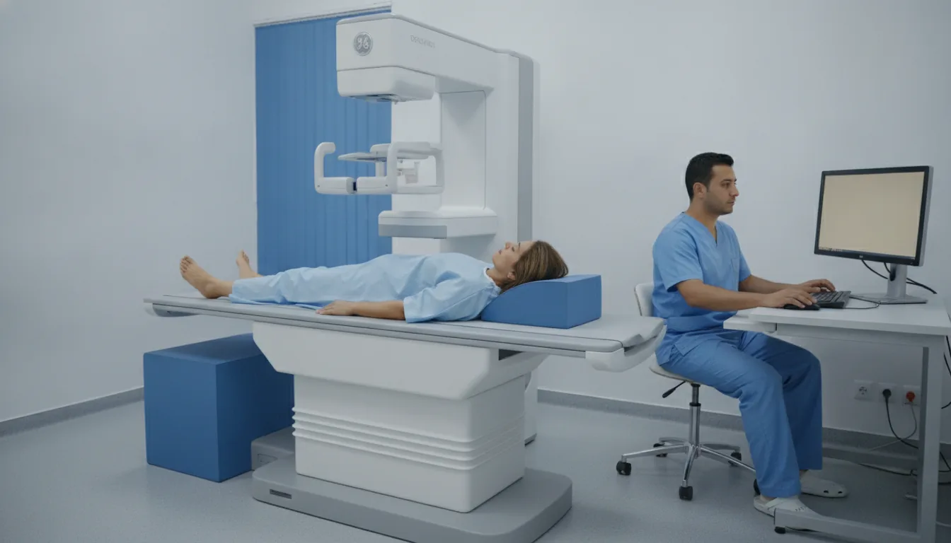
The patient lies on their back on the examination table

2

For the lumbar spine measurement, the legs are raised on a cube-shaped support

3

For the femur measurement, the foot is held in internal rotation by a support

4

The arm of the machine moves slowly over the area being measured

5

The patient must remain still during acquisition (a few seconds per site)

6

Measurements are taken on the lumbar spine and then on the hip (one or both sides)

7

The examination is completely painless and requires no injection

8

The results are calculated automatically by the machine's software

Duration

10 to 15 minutes

Results

The results (T-score and Z-score) are available immediately after the examination. The radiologist writes a report interpreting the values according to the clinical context. The results are sent to the referring physician within 24 to 48 hours.

Risks and side effects

Extremely low radiation dose (less than 0.01 mSv, lower than a chest X-ray)
Completely painless and non-invasive examination
No side effects
Contraindication in case of pregnancy (precaution)
Results may be distorted by the presence of fixation hardware, aortic calcifications or severe osteoarthritis

These risks remain rare. Your physician will inform you in detail before the examination.

Book an Appointment

Contact us to schedule your examination or request a quote.

Request a Quote +216 36 402 000

At a glance

Duration: 10 to 15 minutes
Preparation: Required
Book an appointment