
Surgical biopsies

Surgical biopsies

Surgical sampling of suspicious tissue when simple biopsies are impossible or insufficient, to establish a precise diagnosis.

What is it?

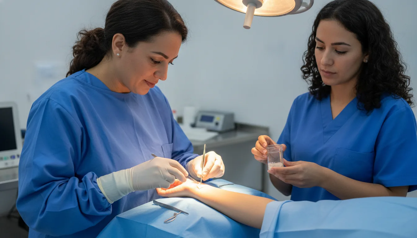
A surgical biopsy consists of removing, in the operating theatre, a fragment or the whole of a suspicious lesion in order to establish its exact nature. It is offered when simpler sampling — needle aspiration or image-guided biopsy — is impossible, insufficient or inconsistent with the images. Depending on the location, the procedure may be an excisional biopsy, which removes the whole lesion (lymph node, subcutaneous nodule, breast lesion), or an incisional biopsy, which takes only a representative fragment. Prior radiological localisation is sometimes performed to guide the surgeon precisely to a non-palpable lesion. At Clinique Pasteur Tunis, surgical biopsies are performed as day surgery in most cases, under appropriate anaesthesia. Pathological analysis of the samples is carried out, with the possibility of frozen-section examination during the procedure when the situation warrants it, and the results are passed on quickly to the doctor to organise the next stage of care.
Surgical biopsies

When is it indicated?

Enlarged lymph node whose nature needs to be determined

A lymph node that remains enlarged without explanation deserves to be analysed: removing it in the operating theatre, a simple procedure, reveals its exact nature.

Suspicious lesion inaccessible to image-guided needle sampling

Some lesions are located in areas the needle cannot reach safely: surgical sampling then becomes the best option.

Previous samples insufficient or inconsistent with the imaging

When the first biopsies were inconclusive or do not match the images, a more complete surgical sample resolves the doubt.

Nodule or soft-tissue mass to be removed for complete analysis

Removing the whole lesion makes it possible both to treat it and to analyse it in full, for as precise a diagnosis as possible.

Non-palpable breast lesion requiring localised removal

For a breast abnormality visible only on the images, radiological localisation guides the surgeon precisely to the area to be removed.

Preparation

1Imaging work-up precisely locating the lesion
2Pre-operative radiological localisation if the lesion is not palpable
3Anaesthesia consultation
4Fast for 6 hours before the procedure if general anaesthesia is planned
5Adjustment of anticoagulants according to medical advice

Procedure

1

Local, regional or general anaesthesia depending on the location

2

Discreet incision over the lesion

3

Removal of the fragment or complete excision of the lesion

4

Frozen-section examination during the procedure if necessary

5

Check for absence of bleeding and careful closure

6

Discharge the same day in most cases

Duration

30 minutes to 1 hour 30

Aftercare & Recovery

The sample undergoes complete pathological analysis, and the results are passed on to the doctor within the following days. This precise diagnosis makes it possible to guide the most appropriate care. The scar is generally discreet and recovery straightforward.

Risks and side effects

Small haematoma or swelling of the sampled area
Local infection, rare and simply treated
Temporary tenderness of the scar

These risks remain rare. Your surgeon will inform you in detail before the procedure.

Request a Quote

Contact us to schedule a consultation or receive a personalized quote.

Request a Quote +216 36 402 000

At a glance

Duration: 30 minutes to 1 hour 30
Preparation: Required
Dedicated specialist team