
General and specialized ultrasound
Centres/Radiology/Ultrasound

General and specialized ultrasound

Imaging technique using ultrasound waves to explore organs and tissues in real time, without ionizing radiation, suitable for many diagnostic indications.

What is it?

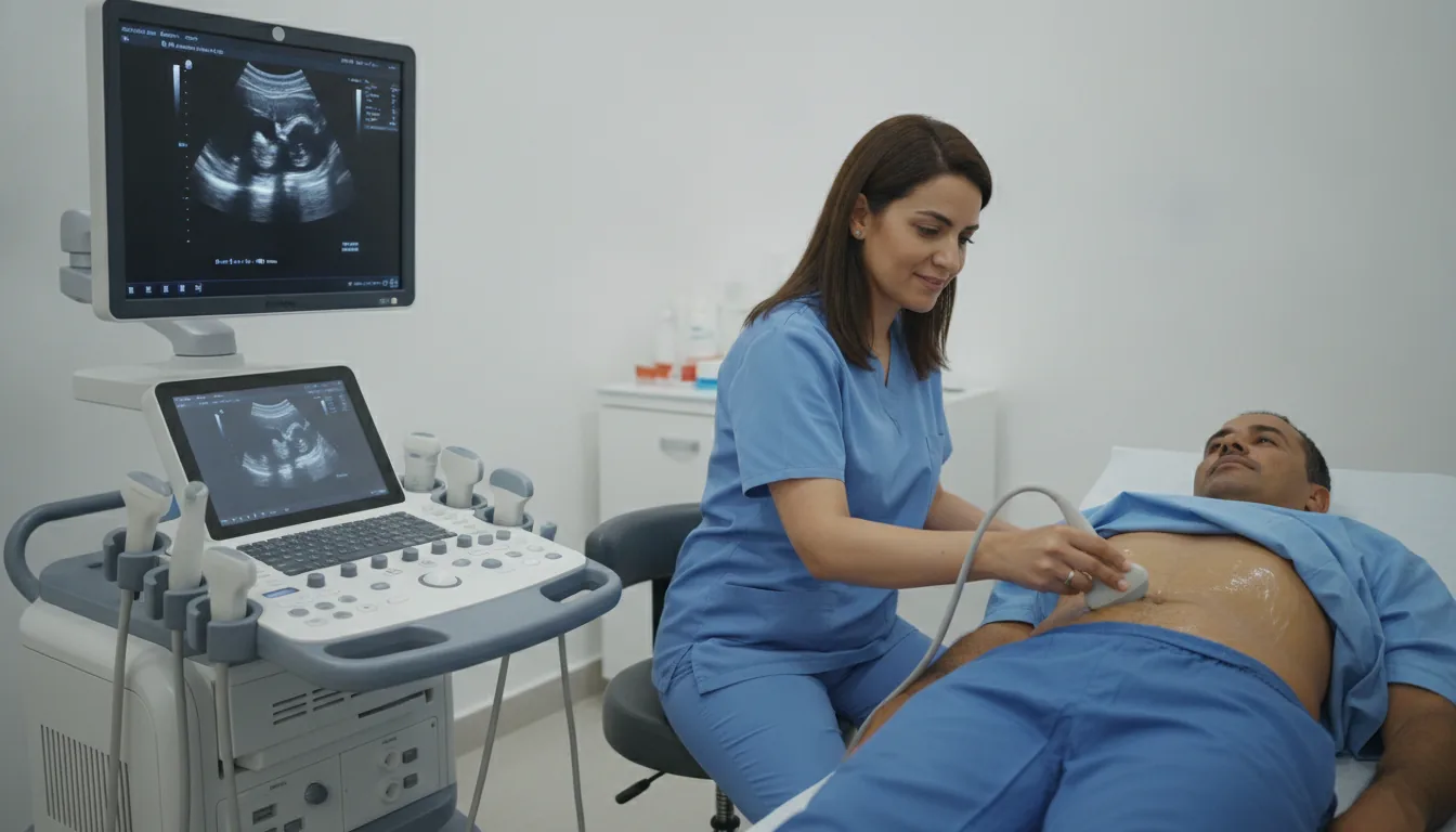
Ultrasound is a medical imaging technique that uses ultrasound waves (high-frequency sound waves) to produce real-time images of the body's internal organs and structures. A transmitting-receiving probe is applied to the skin, sends out ultrasound waves that are reflected differently depending on the nature of the tissues they pass through, and captures the returning echoes to convert them into dynamic images on a screen. Ultrasound is a first-line examination in many clinical situations because of its many advantages: total absence of ionizing radiation, non-invasive and painless nature, ability to examine in real time with dynamic visualization of the organs, reproducibility, availability and moderate cost. It can be repeated without risk, including in pregnant women and children. The fields of application of ultrasound are extremely varied: abdominal ultrasound (liver, gallbladder, pancreas, spleen, kidneys), pelvic and endovaginal ultrasound (uterus, ovaries, prostate), thyroid ultrasound, soft tissue ultrasound (muscles, tendons, lymph nodes), breast ultrasound as a complement to mammography, vascular ultrasound (Doppler of the arteries and veins), and obstetric ultrasound for pregnancy monitoring. Doppler mode also makes it possible to analyze blood flow in real time. At Clinique Pasteur in Tunis, ultrasound examinations are performed on latest-generation machines offering exceptional image resolution. The specialized radiologists adapt the type of probe and the protocol to each indication, guaranteeing a complete examination and an accurate diagnosis.
General and specialized ultrasound

When is it indicated?

Abdominal pain: exploration of the liver, gallbladder, pancreas and kidneys

For abdominal pain, ultrasound is often the first examination requested: it painlessly visualizes the main organs of the abdomen and looks, for example, for gallstones or an abnormality of the liver or kidneys.

Thyroid conditions: nodules, goiter, thyroiditis

If the thyroid gland appears enlarged or if a small lump is felt in the neck, ultrasound determines its size and appearance and helps decide on the appropriate course of action.

Breast conditions: complement to mammography, biopsy guidance

Ultrasound complements mammography, particularly in women with dense breasts. It helps distinguish a fluid-filled cyst from a solid nodule and guides any sampling procedure.

Pregnancy monitoring: ultrasounds of the 1st, 2nd and 3rd trimesters

Throughout pregnancy, ultrasound monitors the baby's growth and well-being in complete safety, without any radiation, for both mother and child.

Pelvic conditions: uterine fibroids, ovarian cysts, prostate disease

To investigate lower abdominal pain, unusually heavy periods or urinary problems, ultrasound examines the uterus, ovaries or prostate simply and painlessly.

Vascular conditions: venous thrombosis, carotid stenosis, aortic aneurysm

Thanks to Doppler mode, ultrasound observes blood flowing through the vessels: it looks for a clot in a swollen leg, checks the arteries of the neck or monitors the diameter of the aorta.

Muscle and tendon conditions: tendinopathies, muscle tears, joint effusions

After exertion or a sports injury, ultrasound examines muscles and tendons in motion, making it possible to confirm a tear or inflammation and adapt the return to activity.

Guidance of interventional procedures: punctures, biopsies, injections

By visualizing the needle in real time, ultrasound allows the doctor to perform samplings or injections with great precision and maximum safety.

Preparation

1Abdominal ultrasound: fast for 6 hours (small amounts of water allowed)
2Pelvic ultrasound: have a full bladder (drink 1 liter of water 1 hour before and do not urinate)
3Thyroid ultrasound: no special preparation
4Obstetric ultrasound of the 1st trimester: half-full bladder
5Soft tissue ultrasound: no preparation
6Vascular ultrasound (Doppler): no special preparation
7Bring the medical prescription and previous examinations
8Wear clothing that is easy to lift or remove over the area to be examined

Procedure

1

The patient is comfortably positioned on the examination table, generally lying on their back

2

Ultrasound gel is applied to the skin over the area to be explored

3

The radiologist applies the probe and moves it gently to visualize the different structures

4

The images appear in real time on the screen

5

The patient may be asked to change position (lying on their side, standing) to optimize the examination

6

Measurements and annotations are made during the examination

7

In Doppler mode, blood flow is analyzed with color coding

8

The gel is wiped off at the end of the examination

Duration

15 to 30 minutes depending on the type of ultrasound

Results

The radiologist communicates the initial results to the patient immediately after the examination. A detailed report with images is written and sent to the referring physician within 24 to 48 hours.

Risks and side effects

No known risk: ultrasound waves emit no ionizing radiation
Completely painless examination (light pressure from the probe)
No known contraindications
Can be repeated without limitation, including during pregnancy
Only limitation: examination quality may be reduced in obese patients or in case of interposed bowel gas

These risks remain rare. Your physician will inform you in detail before the examination.

Book an Appointment

Contact us to schedule your examination or request a quote.

Request a Quote +216 36 402 000

At a glance

Duration: 15 to 30 minutes depending on the type of ultrasound
Preparation: Required
Book an appointment