
Intraoperative neurophysiological monitoring
Centres/Neurophysiological Testing/Intraoperative monitoring

Intraoperative neurophysiological monitoring

Real-time monitoring of nerve function during surgical procedures involving a neurological risk, making it possible to prevent irreversible nerve damage.

What is it?

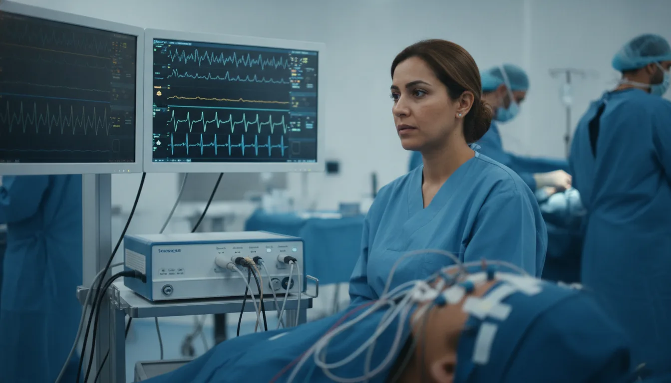
Intraoperative neurophysiological monitoring (IONM) consists of the continuous, real-time monitoring of nerve functions during a surgical procedure. The objective is to detect early any suffering of the nervous system (brain, spinal cord, cranial or peripheral nerves) in order to alert the surgeon and allow an immediate modification of the surgical gesture before an irreversible lesion occurs. The techniques used include somatosensory evoked potentials (SEP), motor evoked potentials (MEP) by transcranial magnetic or electrical stimulation, continuous electromyography (free-running EMG), direct nerve stimulation and electrocorticography. The choice of techniques depends on the type of surgery and the nervous structures at risk. Intraoperative monitoring has considerably improved the safety of spinal surgeries (scoliosis, disc herniation, spinal cord tumours), skull base surgeries, thyroid surgeries (protection of the recurrent nerve) and operations on the peripheral nerves. The presence of a neurophysiologist in the operating theatre has become a quality standard in many procedures involving a neurological risk. The Clinique Pasteur in Tunis offers an intraoperative monitoring service provided by experienced neurophysiologists, working in close collaboration with the surgical and anaesthetic teams to optimise patient safety.
Intraoperative neurophysiological monitoring

When is it indicated?

Surgery for scoliosis and spinal deformities

It makes it possible to monitor the spinal cord during the correction of a spinal deformity, to avoid any lesion.

Surgery for intracanal spinal cord tumours

It helps protect the spinal cord during the removal of a tumour located in the canal of the spine.

Surgery for skull base tumours

It monitors the nearby nerves during a delicate operation at the base of the skull.

Posterior fossa surgery (acoustic neuroma)

It helps preserve the hearing and facial nerves during the removal of a tumour.

Thyroid and parathyroid surgery (monitoring of the recurrent nerve)

It makes it possible to monitor the voice nerve during a thyroid operation, to avoid harming the vocal cords.

Carotid surgery (endarterectomy)

It helps monitor the brain during an operation on the neck artery that supplies it.

Surgery for intracranial aneurysms

It monitors the functioning of the brain in real time during the treatment of a dilatation of a cerebral artery.

Peripheral nerve surgery

It helps protect and identify the nerves during an operation that directly concerns them.

Epilepsy surgery (cortical mapping)

It helps identify the important areas of the brain to be preserved during an epilepsy operation.

Preparation

1The monitoring is planned in advance by the surgeon in consultation with the neurophysiologist.
2A pre-operative neurophysiological assessment (SEP, MEP, EMG) is often carried out as a reference.
3The patient is informed of the neurophysiological monitoring at the pre-operative consultation.
4The anaesthetic protocol is adapted in consultation with the anaesthetist to allow the monitoring (avoiding prolonged muscle relaxants, adjusting the halogenated agents).
5The neurophysiologist reviews the medical file and the type of surgery planned.

Procedure

1

Placement of the stimulation and recording electrodes after the anaesthetic induction.

2

Recording of the reference values (baseline) before the start of the surgical gesture.

3

Continuous monitoring of the neurophysiological parameters throughout the procedure.

4

Real-time communication with the surgeon and the anaesthetist in the event of a significant change in the signals.

5

In the event of an alert (a drop in amplitude > 50 % or an increase in latency > 10 %), the surgeon is immediately notified.

6

Adjustment of the monitoring techniques according to the operative steps and the structures at risk.

7

Checking of the parameters at the end of the procedure before closure.

8

Drafting of a monitoring report detailing the significant events.

Duration

Duration of the surgical procedure (from 2 to 8 hours depending on the type of surgery)

Results

A monitoring report is drawn up and integrated into the operative file. It details the techniques used, the reference values, any alerts and the corrective measures taken. This document is sent to the surgeon and the anaesthetist.

Risks and side effects

Skin irritation at the electrode fixation points.
Exceptional superficial burn at the level of the stimulation electrodes.
Possible tongue bite during transcranial motor stimulation (a mouth guard is systematically put in place).

These risks remain rare. Your physician will inform you in detail before the examination.

Book an Appointment

Contact us to schedule your examination or request a quote.

Request a Quote +216 36 402 000

At a glance

Duration: Duration of the surgical procedure (from 2 to 8 hours depending on the type of surgery)
Preparation: Required
Book an appointment