
Endoscopic mucosal resection (EMR)
Centres/Endoscopy/Mucosal resection

Endoscopic mucosal resection (EMR)

Advanced endoscopic technique for resecting extensive superficial lesions of the digestive mucosa, a minimally invasive alternative to surgery.

What is it?

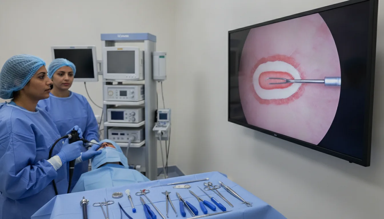
Endoscopic mucosal resection (EMR) is an advanced endoscopic technique that makes it possible to remove large superficial lesions from the lining of the digestive tract. Unlike standard polypectomy, EMR is suited to flat or sessile lesions larger than 20 mm, laterally spreading adenomas and superficial cancers that do not extend beyond the submucosa. The technique is based on injecting a solution into the submucosa (submucosal cushion) in order to lift the lesion and separate it from the deep layers of the digestive wall. This lifting allows a safer resection by reducing the risk of perforation and making it easier to capture the lesion with the diathermy snare. The resection can be performed in a single piece (en bloc) or in several fragments (piecemeal) depending on the size of the lesion. Histological analysis of the resected specimens is fundamental: it confirms the nature of the lesion, assesses the resection margins and determines whether the endoscopic treatment is sufficient or whether additional surgical management is necessary. EMR thus makes it possible to avoid major surgery in many patients. At Clinique Pasteur in Tunis, EMR procedures are performed by endoscopists experienced in advanced resection techniques. The procedure takes place under general anesthesia in a safe environment, with close follow-up to prevent complications.
Endoscopic mucosal resection (EMR)

When is it indicated?

Large flat or sessile adenomas (larger than 20 mm)

Broad, flat polyps cannot be removed with a simple snare. EMR makes it possible to remove them safely, by first lifting them with an injection beneath the mucosa.

Laterally spreading tumors (LST) of the colon

Some lesions spread across the surface of the colon wall rather than forming a lump. This specialized technique makes it possible to remove them completely despite their extent.

Superficial cancers of the digestive tract confined to the mucosa

When a cancerous lesion is detected very early and remains confined to the superficial layer of the wall, it can often be removed entirely endoscopically, without surgery.

Dysplastic lesions in Barrett's esophagus

In people whose esophageal lining has been transformed by reflux, areas that are beginning to change can be removed preventively using this technique.

Large gastric polyps

Large stomach polyps can be removed by EMR, which makes it possible both to analyze them completely and to avoid stomach surgery.

Lesions not amenable to standard polypectomy

Some lesions are poorly located or awkwardly shaped for standard removal. EMR offers an additional solution for removing them without resorting to surgery.

Alternative to surgery for extensive benign lesions

For many benign but extensive lesions, this technique avoids major surgery: the hospital stay is shorter and recovery considerably faster.

Preparation

1Same preparation as for colonoscopy for colonic lesions
2Fast for at least 8 hours for upper digestive tract lesions
3Discontinuation of anticoagulants and antiplatelet agents according to the prescribed protocol (5 to 10 days before)
4A recent coagulation panel is mandatory (PT, aPTT, platelets)
5Mandatory pre-anesthesia consultation
6Hospitalization for 24 to 48 hours is often necessary
7Bring previous endoscopy reports and biopsy results

Procedure

1

General anesthesia is induced by the anesthesiologist

2

The lesion is located and its margins are delineated (chromoendoscopy)

3

A solution is injected into the submucosa to lift the lesion

4

Proper lifting of the lesion is verified (positive lifting sign)

5

The diathermy snare is positioned around the lifted lesion

6

The resection is performed by applying an electric current

7

The fragments are retrieved for histological analysis

8

The resection site is examined and hemostatic measures are applied (clips, coagulation)

9

The patient is hospitalized for monitoring for 24 to 48 hours

Duration

30 to 90 minutes depending on the size and location of the lesion

Results

The gastroenterologist communicates the initial visual findings after the examination. The final results of the histological analysis are available within 10 to 21 days and are discussed during a follow-up consultation.

Risks and side effects

Immediate or delayed bleeding (5 to 10 % of cases, usually controlled endoscopically)
Digestive perforation (1 to 2 % of cases)
Incomplete resection requiring a second procedure or surgery
Local recurrence in case of piecemeal resection (10 to 20 %, monitored by endoscopy)
Post-resection syndrome (pain, fever)
Late scar stricture (for circumferential lesions)

These risks remain rare. Your physician will inform you in detail before the examination.

Book an Appointment

Contact us to schedule your examination or request a quote.

Request a Quote +216 36 402 000

At a glance

Duration: 30 to 90 minutes depending on the size and location of the lesion
Preparation: Required
Book an appointment