
Gastroscopy (Upper GI Endoscopy)
Centres/Endoscopy/Gastroscopy

Gastroscopy (Upper GI Endoscopy)

Visual examination of the esophagus, stomach and duodenum using a flexible endoscope, allowing the diagnosis of upper digestive tract conditions.

What is it?

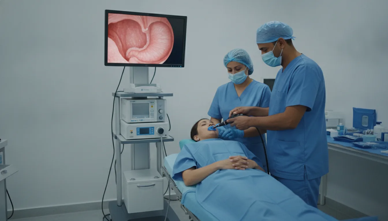
Upper gastrointestinal endoscopy (gastroscopy, also known as OGD) is the gold-standard examination for exploring the upper part of the digestive tract. It provides direct visualization of the lining of the esophagus, stomach and duodenum using a flexible endoscope fitted with a miniaturized camera and a light source. This examination is essential for diagnosing many digestive conditions. Gastroscopy can detect and diagnose gastric and duodenal ulcers, gastritis, esophagitis, gastroesophageal reflux disease, hiatal hernias, polyps, benign or malignant tumors, as well as esophageal varices. During the examination, the gastroenterologist can take biopsies (tissue samples) for histological analysis, in particular to test for Helicobacter pylori. The examination is generally well tolerated thanks to local anesthesia of the throat with a spray or, if the patient prefers, under light sedation. The examination itself is short, around 5 to 10 minutes, and the visual findings are communicated immediately by the physician. At Clinique Pasteur in Tunis, gastroscopy is performed by experienced gastroenterologists using latest-generation endoscopes offering high-definition image quality. The equipment is rigorously disinfected according to current protocols, guaranteeing optimal safety for every patient.
Gastroscopy (Upper GI Endoscopy)

When is it indicated?

Persistent or recurrent upper abdominal pain

When pain in the pit of the stomach keeps coming back despite treatment, gastroscopy allows the physician to look directly at the lining to find the cause, such as gastritis or an ulcer.

Heartburn or gastroesophageal reflux resistant to treatment

If acid reflux persists despite medication, the examination checks the condition of the esophagus, looks for inflammation or a hiatal hernia, and helps adjust the treatment.

Difficulty swallowing (dysphagia)

Discomfort or a blocking sensation when swallowing always deserves investigation: gastroscopy visualizes the esophagus to identify a narrowing, inflammation or any other abnormality.

Unexplained nausea or vomiting

When nausea or vomiting persists without explanation, examining the stomach and duodenum makes it possible to look for a local cause and guide treatment.

Unexplained iron-deficiency anemia

Iron deficiency can result from small, silent bleeding in the digestive tract. Gastroscopy looks for a lesion of the esophagus, stomach or duodenum that could be responsible.

Testing for Helicobacter pylori

This very common bacterium can cause gastritis and ulcers. Small, painless samples taken during the examination make it possible to detect and treat it effectively.

Surveillance of pre-cancerous lesions (Barrett's esophagus, polyps)

Some benign lesions require regular monitoring to make sure they are not progressing. Scheduled check-ups make it possible to act very early if any change occurs.

Upper gastrointestinal bleeding

In case of vomiting blood or very dark stools, gastroscopy quickly locates the source of the bleeding and often allows it to be treated during the same procedure.

Preparation

1Fast strictly for at least 6 hours before the examination (no eating, drinking or smoking)
2Report any ongoing anticoagulant or antiplatelet treatment
3Report any known allergies, particularly to local anesthetics
4Bring the results of previous examinations (X-rays, blood tests)
5Arrange for someone to accompany you if the examination is performed under sedation
6Do not drive for 12 hours after sedation

Procedure

1

The patient lies down on their left side

2

Local anesthesia of the throat is applied with a spray, or light sedation is administered intravenously

3

A mouthpiece is placed between the teeth to protect the endoscope

4

The flexible endoscope is gently inserted through the mouth and guided down to the duodenum

5

The physician carefully examines the lining of the esophagus, stomach and duodenum

6

Biopsies may be taken if necessary (painless)

7

The endoscope is gently withdrawn at the end of the examination

8

The patient is monitored in a recovery room for 30 minutes to 1 hour

Duration

5 to 15 minutes for the examination, 1 to 2 hours including preparation and monitoring

Results

The visual findings are communicated immediately after the examination by the gastroenterologist. Biopsy results are available within 5 to 10 working days.

Risks and side effects

Mild throat discomfort or soreness for 24 to 48 hours after the examination
Minor bleeding at a biopsy site (rare and usually harmless)
Digestive perforation (exceptional, less than 1/10,000)
Reaction to the anesthesia or sedation (rare)
Infection (extremely rare thanks to strict disinfection protocols)

These risks remain rare. Your physician will inform you in detail before the examination.

Book an Appointment

Contact us to schedule your examination or request a quote.

Request a Quote +216 36 402 000

At a glance

Duration: 5 to 15 minutes for the examination, 1 to 2 hours including preparation and monitoring
Preparation: Required
Book an appointment