
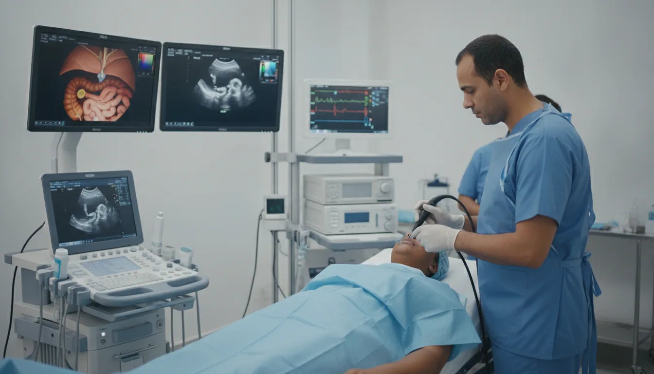
Endoscopic Ultrasound (EUS)
Examination combining endoscopy and ultrasound to obtain precise images of the digestive walls and adjacent organs, essential for the assessment of certain digestive and pancreatic conditions.
What is it?
When is it indicated?
By placing the ultrasound probe in direct contact with the lesion, this examination precisely measures the depth of a tumor within the digestive wall, essential information for choosing the best treatment.
The pancreas is a deep organ that is difficult to examine from the outside. EUS gets as close to it as possible and can detect very small lesions invisible on other examinations.
When a stone or an obstruction is suspected in the ducts that carry bile, this highly sensitive examination can confirm or rule it out before considering a therapeutic procedure.
Some bumps seen during a standard endoscopy develop beneath the surface of the wall. Internal ultrasound shows which layer they arise from and helps clarify their nature.
Under ultrasound guidance, a very fine needle can sample a few cells from a suspicious lesion or lymph node. This sampling provides a precise diagnosis while avoiding surgery.
When facing a fluid-filled pocket in the pancreas, the examination studies its contents and walls to distinguish harmless cysts from those requiring surveillance or treatment.
In case of prolonged inflammation of the pancreas, EUS finely assesses the condition of the gland and its ducts, which helps monitor the disease and adjust its management.
Some pockets of fluid that form around the pancreas can be drained directly through the stomach wall, under ultrasound guidance, thereby avoiding more invasive surgery.
Preparation
Procedure
Deep sedation is administered by the anesthesiologist
The echoendoscope is introduced through the mouth (upper route) or through the anus (lower route, depending on the indication)
The endoscope is positioned facing the area to be explored
The ultrasound probe produces real-time images of the digestive wall and adjacent organs
The physician analyzes the size, structure and extension of the lesions
If indicated, a fine-needle aspiration is performed under ultrasound guidance
The samples are sent to the laboratory for analysis
The patient is monitored in the recovery room for 1 to 2 hours
Duration
30 to 60 minutes for the examination, 2 to 3 hours in total
Results
The imaging findings are communicated by the gastroenterologist in the days following the examination. The results of needle aspirations are available within 7 to 14 days.
Risks and side effects
These risks remain rare. Your physician will inform you in detail before the examination.
Book an Appointment
Contact us to schedule your examination or request a quote.
Request a Quote +216 36 402 000