
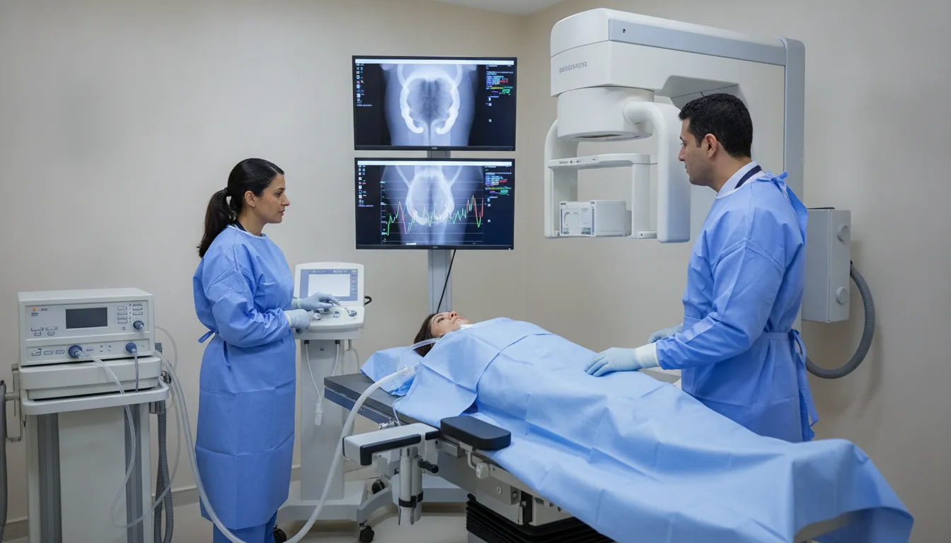
Video-urodynamics
Combination of a urodynamic assessment and real-time radiological imaging to visualise bladder morphology and function simultaneously.
What is it?
When is it prescribed?
It provides a very complete evaluation of both the shape and the function of the bladder in neurological patients.
It helps detect an abnormal backflow of urine from the bladder towards the kidneys, which is potentially harmful.
It is useful when the bladder and the sphincter do not coordinate properly during voiding.
It helps investigate abnormalities of the urinary tract present from birth.
It is offered when an operation for urinary leakage has not given the expected result, to understand why.
It provides additional information when the usual tests have been inconclusive.
It helps check the proper functioning of a surgical construction created to drain urine.
Preparation
How the exam is performed
Installation in a radiology room equipped with a fluoroscopy unit
Insertion of the bladder catheter and the rectal pressure sensor
Filling of the bladder with a warm radio-opaque contrast agent
Simultaneous recording of pressures and radiological images
Images taken at different stages of filling and during voiding
Real-time detection of possible vesicoureteral reflux
Voiding phase with simultaneous video and manometric recording
Removal of equipment and post-void images if necessary
Duration
45 minutes to 1 hour 15 minutes
Results
The report combines manometric data (pressures, flows, compliance) and morphological data (fluoroscopic images). It can detect vesicoureteral reflux, dyssynergia and anatomical abnormalities and correlate them with the functional data. Results available within 48 to 72 hours.
Risks and side effects
These risks remain rare. Your physician will explain them in detail before the exam.
Book an Appointment
Contact us to schedule your Video-urodynamics or request a quote.
Request a Quote +216 36 402 000