
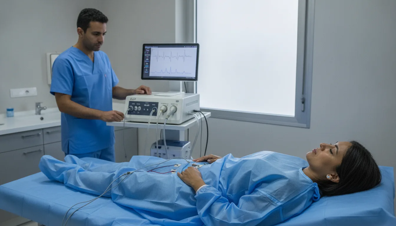
Perineal electromyography
Recording of the electrical activity of the pelvic floor muscles and the urethral sphincter to assess their innervation and coordination.
What is it?
When is it prescribed?
It helps identify a lack of coordination between the bladder and the sphincter when urinating.
It evaluates the muscles of the perineum after childbirth or an operation that may have affected their nerves.
It is useful when urinary leakage could be linked to a problem with the nerves of the perineum.
It helps assess the muscles of the perineum before starting an appropriate rehabilitation programme.
It helps explain difficulty emptying the bladder related to the nervous system.
It helps investigate damage to the nerve of the perineum that can cause pain or control problems.
It monitors the coordination of the perineal muscles in patients whose bladder is disturbed by a neurological disease.
Preparation
How the exam is performed
Installation in the gynaecological position or lying on the side
Placement of surface electrodes on the perineum or insertion of needle electrodes into the target muscles
Recording of muscle activity at rest
Recording during voluntary contraction of the perineum
Recording during straining and coughing efforts
Assessment of sphincter coordination during bladder filling (if combined with the urodynamic assessment)
Removal of the electrodes at the end of the test
Duration
20 to 30 minutes (up to 45 minutes if combined with the urodynamic assessment)
Results
The analysis covers muscle activity at rest, the quality of recruitment during voluntary contraction, the presence of denervation potentials and sphincter coordination during the voiding cycle. The report is available within 48 hours.
Risks and side effects
These risks remain rare. Your physician will explain them in detail before the exam.
Book an Appointment
Contact us to schedule your Perineal EMG or request a quote.
Request a Quote +216 36 402 000