
Visual evoked potentials (VEP)

Visual evoked potentials (VEP)

Recording of the electrical response of the visual cortex to a light stimulation, making it possible to assess the integrity of the visual pathways from the optic nerve to the occipital cortex.

What is it?

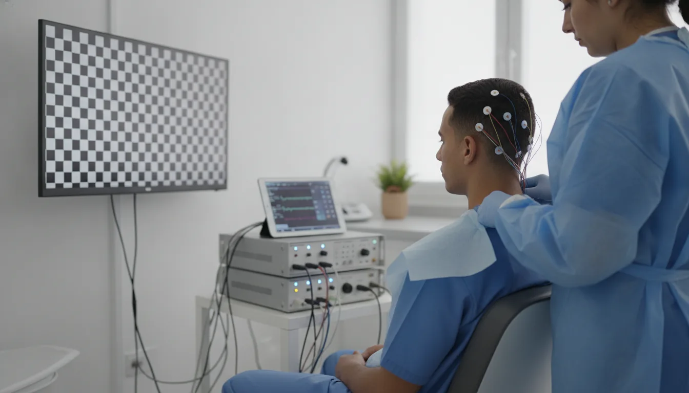
Visual evoked potentials (VEP) are a neurophysiological examination that measures the electrical response of the occipital visual cortex to a standardised visual stimulation. The examination consists of recording, using electrodes placed on the scalp over the occipital cortex, the electrical signals generated by the brain in response to a visual stimulus, most often an alternating checkerboard displayed on a screen. This examination explores the functional integrity of the entire visual pathways, from the retina to the occipital cortex, passing through the optic nerve, the optic chiasm and the optic radiations. The main wave analysed is the P100 wave, whose latency (time of appearance) and amplitude are measured. A lengthening of the P100 wave latency is a very sensitive marker of demyelination of the optic nerve. VEP have a privileged place in the diagnosis of multiple sclerosis, where they make it possible to detect optic neuritis even when clinically silent. A unilateral lengthening of the P100 latency strongly suggests retrobulbar optic neuritis, whether symptomatic or subclinical. The Clinique Pasteur in Tunis carries out VEP according to the standardised protocols of the International Federation of Clinical Neurophysiology (IFCN), ensuring reliable results that are comparable over time.
Visual evoked potentials (VEP)

When is it indicated?

Suspected multiple sclerosis (looking for subclinical optic neuritis)

It helps detect a condition of the optic nerve, sometimes without symptoms, as part of the assessment of multiple sclerosis.

Retrobulbar optic neuritis

It is useful for exploring an inflammation of the optic nerve responsible for a drop in vision.

Optic neuropathy (compressive, toxic, ischaemic, hereditary)

It makes it possible to assess the functioning of the optic nerve when it is affected for various reasons.

Unexplained drop in visual acuity

It helps specify the origin of a reduction in vision whose cause has not yet been established.

Tumours compressing the visual pathways (pituitary adenoma, meningioma)

It provides information when a mass may be pressing on the visual pathways.

Monitoring of demyelinating conditions

It makes it possible to follow the course of diseases affecting the protective sheath of the nerves, including those of vision.

Functional assessment of the visual pathways in uncooperative children

It offers a way to objectively assess vision in a child who cannot cooperate with a conventional examination.

Assessment of neurosarcoidosis or Behçet's disease with neurological involvement

It helps explore the impact of these inflammatory diseases when they affect the nervous system.

Preparation

1Bring your usual corrective glasses or contact lenses (visual acuity must be optimal).
2Wash your hair without styling products.
3The examination does not require fasting.
4Report any known ophthalmological condition (cataract, glaucoma, macular degeneration).
5Report the taking of any medication that may affect vision.
6Bring the results of recent ophthalmological and neurological examinations.

Procedure

1

The patient is settled comfortably seated in front of a screen, at a standardised distance.

2

Placement of electrodes on the scalp over the occipital visual cortex.

3

The examination is carried out eye by eye: a patch is placed alternately over each eye.

4

The patient must fix a central point on the screen displaying a black-and-white checkerboard whose squares reverse at a regular frequency.

5

The computer averages the cortical responses over 100 to 200 stimulations to extract the VEP signal from the background noise.

6

Measurement of the latency and amplitude of the P100 wave for each eye.

7

In the event of poor visual acuity, stimulation by a flash of light may be used.

Duration

30 to 45 minutes

Results

The neurophysiologist analyses the tracings and compares the latencies and amplitudes with the normative values. The results are available within 24 to 48 hours. A lengthening of the P100 latency or a significant inter-ocular asymmetry are the main abnormalities looked for.

Book an Appointment

Contact us to schedule your examination or request a quote.

Request a Quote +216 36 402 000

At a glance

Duration: 30 to 45 minutes
Preparation: Required
Non-invasive examination
Book an appointment