
Stress echocardiography

Stress echocardiography

Echocardiography performed during physical exercise or under pharmacological stimulation, to detect abnormalities of heart contraction revealed by stress.

What is it?

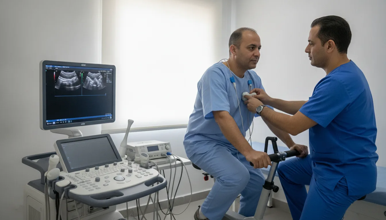
Stress echocardiography is an examination that combines ultrasound imaging of the heart with stress, whether physical (exercise on a semi-recumbent bicycle or treadmill) or pharmacological (intravenous injection of dobutamine). The aim is to compare how the heart works at rest and under stress, in order to detect contraction abnormalities that are only visible when the heart is subjected to an increased workload. This examination is an essential diagnostic tool for detecting coronary artery disease. When a coronary artery is narrowed, the area of heart muscle it supplies may contract normally at rest but show a contraction abnormality during exercise, because the oxygen supply becomes insufficient. Stress echocardiography detects these segmental wall motion abnormalities with excellent sensitivity and specificity. Pharmacological stress echocardiography with dobutamine is particularly useful in patients who cannot perform sufficient physical exercise (joint limitations, respiratory failure, peripheral artery disease of the lower limbs). Dobutamine is a medication that stimulates the heart in a way comparable to physical exercise. At the Clinique Pasteur in Tunis, stress echocardiography is performed by experienced cardiologists in a secure environment. The examination is continuously monitored, with resuscitation equipment immediately available, ensuring optimal patient safety.
Stress echocardiography

When is it indicated?

Suspected coronary artery disease in a patient unable to perform maximal exercise

When joint or breathing problems prevent running or pedaling hard enough, a medication reproduces the effect of exercise to test the heart gently.

Uninterpretable or inconclusive exercise ECG

If the standard stress test has not given a clear answer, adding ultrasound images shows directly how the heart contracts and settles the question.

Assessment of myocardial viability after infarction

After a heart attack, the examination determines whether the affected areas of heart muscle are still alive and could recover after revascularization treatment.

Preoperative assessment in a patient at cardiovascular risk

Before major surgery, this test checks that the heart will withstand the stress of the operation in a person with risk factors.

Assessment of the severity of valve disease during exercise

Some valve diseases only truly reveal themselves during exercise: the examination observes how the valve behaves when the heart speeds up.

Follow-up after coronary revascularization

After stent placement or bypass surgery, this test checks that blood is once again flowing properly through the heart muscle during exercise.

Assessment of myocardial contractile reserve

The examination measures the heart's ability to increase its contraction strength when challenged, a valuable indicator of its state of health.

Preparation

1Fast for at least 4 hours before the examination
2Report all current medications, especially beta-blockers
3The cardiologist may ask you to stop beta-blockers 48 hours before the examination
4Avoid caffeine and theophylline in the 24 hours before the examination
5Wear comfortable clothing if physical exercise is planned
6Report any history of allergy
7Bring the results of previous cardiology examinations

Procedure

1

A resting ECG and a baseline echocardiogram are performed

2

An intravenous line is placed (for pharmacological stress echo)

3

For physical stress: the patient pedals on a semi-recumbent bicycle or walks on a treadmill

4

For pharmacological stress: dobutamine is infused at progressively increasing doses

5

Ultrasound images are recorded at each stress level

6

The ECG and blood pressure are monitored continuously

7

The examination continues until the target heart rate is reached or positive signs appear

8

The images at rest and at peak stress are compared side by side

9

A recovery phase is monitored

Duration

45 minutes to 1 hour

Results

Results are communicated by the cardiologist after analysis of the images. A detailed report is provided the same day or the following day.

Risks and side effects

Palpitations and increased heart rate (expected effect)
Chest pain (a sign being looked for, leading to stopping the examination)
Heart rhythm disturbance (rare, managed immediately)
Low blood pressure or a drop in blood pressure (rare)
Nausea or headache with dobutamine (temporary)
Risk of a serious cardiac event (exceptional, less than 1/5000)

These risks remain rare. Your physician will inform you in detail before the examination.

Book an Appointment

Contact us to schedule your examination or request a quote.

Request a Quote +216 36 402 000

At a glance

Duration: 45 minutes to 1 hour
Preparation: Required
Book an appointment