
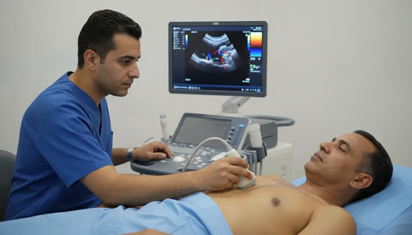
Transthoracic echocardiography (TTE)
Ultrasound imaging of the heart through the chest wall, used to assess the structure and function of the heart chambers, valves and pericardium.
What is it?
When is it indicated?
When the doctor hears an unusual sound through the stethoscope, the ultrasound shows the heart valves and determines whether the murmur is harmless or linked to an abnormality.
If the heart seems to lack strength or if breathlessness persists with no obvious cause, the examination directly measures the heart's ability to pump blood.
When chest pain occurs, the ultrasound helps check that the heart muscle is contracting normally and guides the diagnosis.
After a heart attack, the examination assesses the areas of the heart that may have been affected and checks that pumping strength remains satisfactory.
When a heart valve leaks or narrows, regular ultrasound scans track its progression and help choose the right time for any intervention.
Long-standing high blood pressure can thicken and tire the heart muscle: the ultrasound checks whether the heart shows any signs of this.
In cases of heart muscle disease, the examination specifies the shape, thickness and function of the heart to guide treatment.
In children and adults alike, ultrasound can detect a heart malformation present from birth, completely painlessly.
Before certain operations, the examination makes sure the heart is fit to withstand the anesthesia and the planned procedure.
If an infection of the heart valves is suspected, particularly in cases of persistent fever, the ultrasound looks for signs of valve involvement.
Preparation
Procedure
The patient lies on their back and then on their left side on the examination table
A conductive gel is applied to the chest to help transmit the ultrasound waves
The cardiologist moves the probe over different areas of the chest (acoustic windows)
Images of the heart are recorded from different angles and in different modes
Doppler measurements of blood flow are taken
The patient may be asked to change position or breathing during the examination
The gel is wiped off at the end of the examination
Duration
20 to 40 minutes
Results
Results are available immediately. The cardiologist writes a detailed report with the measurements and conclusions, given to the patient the same day or sent to the prescribing physician.
Book an Appointment
Contact us to schedule your examination or request a quote.
Request a Quote +216 36 402 000