
Somatosensory evoked potentials (SEP)

Somatosensory evoked potentials (SEP)

Recording of the electrical responses of the sensory pathways from the peripheral nerves to the cerebral cortex, assessing the integrity of the lemniscal pathway.

What is it?

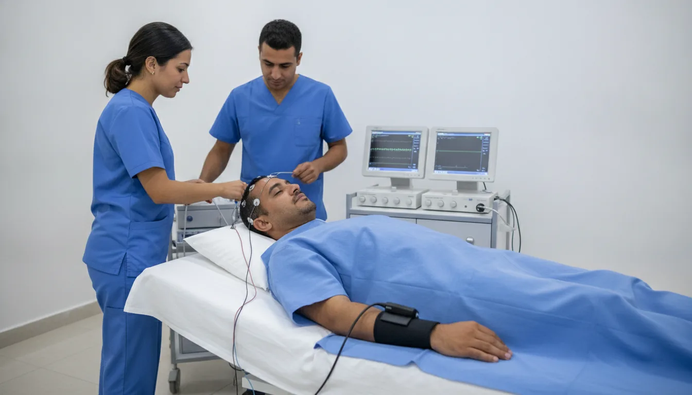
Somatosensory evoked potentials (SEP) explore the lemniscal sensory pathway (the pathway of deep sensation and fine touch) in its entirety, from the peripheral nerves to the parietal cortex. The examination consists of electrically stimulating a peripheral nerve (usually the median nerve at the wrist or the posterior tibial nerve at the ankle) and recording the responses at different levels along the sensory pathways. The upper-limb SEP (median nerve stimulation) record responses at the level of the brachial plexus (Erb's point), the cervical spinal cord, the brainstem and the parietal cortex. The lower-limb SEP (posterior tibial nerve stimulation) explore the sensory pathways from the cauda equina to the parietal cortex, passing through the spinal cord. This examination is particularly indicated in the assessment of cervical and thoracic myelopathies, spinal cord compressions, multiple sclerosis and brainstem lesions. It is also used intraoperatively for the monitoring of the spinal cord during high-risk spinal surgeries. The Clinique Pasteur in Tunis carries out SEP according to international recommendations, allowing a complete and reliable exploration of the somatosensory pathways. The examination is often coupled with VEP and AEP to make up a multimodal assessment of the evoked potentials.
Somatosensory evoked potentials (SEP)

When is it prescribed?

Multiple sclerosis (looking for subclinical involvement of the sensory pathways)

It helps identify a subtle condition of the sensory pathways as part of the assessment of multiple sclerosis.

Cervical spondylotic myelopathy (cervical spinal cord compression)

It is useful when wear of the neck vertebrae compresses the spinal cord, to assess its impact.

Spinal cord compression of another origin (tumoral, disc herniation)

It makes it possible to assess the functioning of the spinal cord when it is compressed by a herniation or a mass.

Traumatic spinal cord lesions

It helps assess the sensory pathways after an injury to the spinal cord.

Cauda equina syndrome

It is useful for exploring the involvement of the nerves at the bottom of the spine, causing sensory disturbances.

Brainstem conditions

It provides information on the sensory pathways passing through a deep region of the brain.

Intraoperative monitoring during spinal surgeries

It makes it possible to monitor the spinal cord in real time during a spinal operation, to protect it.

Prognostic assessment in coma and severe head trauma

It helps the medical team assess the state of the brain in seriously affected patients.

Inflammatory or infectious myelitis

It is useful for exploring an inflammation or infection of the spinal cord.

Preparation

1No fasting is required.
2Wash your hair without styling products.
3Keep the limbs warm to facilitate nerve stimulation.
4Report any history of sensory disturbances, neuropathic pain or spinal cord condition.
5Bring the results of spinal and brain MRIs if available.
6Bring the prescription and the list of current treatments.

How the exam is performed

1

The patient is settled in a semi-reclined position, relaxed.

2

Placement of recording electrodes on the scalp (parietal cortex), the nape of the neck (cervical spinal cord) and the supraclavicular fossa (Erb's point) or the lumbar region.

3

Electrical stimulation of the median nerve at the wrist (upper limbs) or of the posterior tibial nerve at the ankle (lower limbs).

4

The stimulation causes a slight twitch of the thumb or foot, painless to moderately unpleasant.

5

The computer averages the responses over 500 to 1,000 stimulations.

6

Analysis of the latencies of the different components at the peripheral, spinal and cortical levels.

7

Calculation of the central conduction times (from the spinal level to the cortex).

Duration

45 minutes to 1 hour

Results

The results are interpreted by the neurophysiologist taking the clinical context into account. The report, available within 48 hours, specifies the conduction times at each level and identifies the possible site of the abnormality.

Risks and side effects

An unpleasant sensation during the electrical stimulations, generally well tolerated.

These risks remain rare. Your physician will explain them in detail before the exam.

Book an Appointment

Contact us to schedule your SEP or request a quote.

Request a Quote +216 36 402 000

At a glance

Duration: 45 minutes to 1 hour
Preparation: Required
Book an appointment