
Needle electromyography (EMG)

Needle electromyography (EMG)

Examination exploring the electrical activity of muscles at rest and during contraction, making it possible to diagnose muscle disorders and peripheral nerve conditions.

What is it?

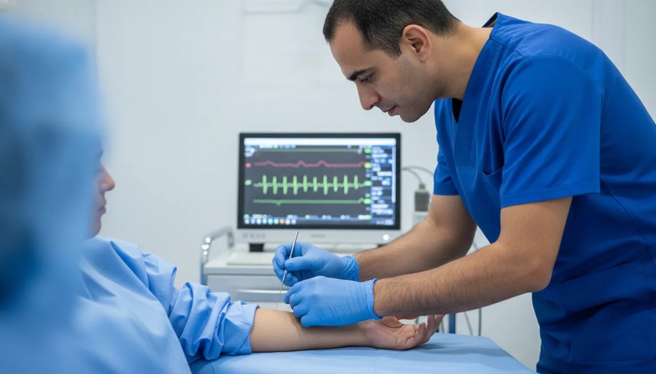
Needle electromyography (EMG) is a neurophysiological examination that analyses the electrical activity of muscles using a fine needle electrode inserted into the muscle. This examination makes it possible to assess the integrity of the motor unit, that is, the whole formed by the motor neuron, its axon and the muscle fibres it innervates. Needle EMG successively explores the activity of the muscle at rest (looking for abnormal spontaneous activity such as fibrillations, fasciculations or myotonic discharges), then during a voluntary contraction effort (analysis of the motor unit potentials: morphology, duration, amplitude, recruitment). These parameters make it possible to distinguish a neurogenic condition (nerve disease) from a myogenic condition (muscle disease). This examination is fundamental in the diagnosis of peripheral neuropathies, radiculopathies (sciatica, cruralgia), motor neuron diseases (amyotrophic lateral sclerosis), myopathies and myositides. It provides information complementary to nerve conduction studies and makes it possible to specify the site, severity and duration of the condition. At the Clinique Pasteur in Tunis, the EMG is performed by experienced neurophysiologists using latest-generation electromyography equipment, allowing a fine and precise analysis of the motor unit potentials.
Needle electromyography (EMG)

When is it prescribed?

Suspected motor neuron disease (amyotrophic lateral sclerosis)

It helps the physician explore a disease affecting the nerves that control the muscles when suggestive signs appear.

Cervical or lumbar radiculopathies (sciatica, cruralgia)

It is useful when faced with pain radiating into the arm or leg, to determine whether a nerve root is compressed.

Peripheral neuropathies (diabetic, alcoholic, inflammatory)

It makes it possible to explore a condition affecting the nerves, for example related to diabetes, and to assess its extent.

Carpal tunnel syndrome and other entrapment syndromes

It helps confirm the compression of a nerve, such as at the wrist, causing tingling or numbness.

Myopathies and muscular dystrophies

It helps distinguish a disease of the muscle itself from a condition of the nerve that controls it.

Myositides and polymyositides

It helps explore an inflammation of the muscles responsible for weakness or pain.

Unexplained muscle weakness

It is offered when a loss of strength with no obvious cause needs to be explored.

Localised or diffuse muscle wasting

It helps understand the origin of muscle wasting, whether it affects a specific area or the whole body.

Persistent fasciculations or muscle cramps

It is useful when faced with repeated and bothersome muscle twitches or cramps.

Preparation

1No fasting is required.
2Come with clean skin, without cream or oil on the areas to be examined.
3Inform the physician if you are taking anticoagulants or antiplatelet agents.
4Report any history of coagulation disorders.
5Bring the prescription, the imaging results (MRI, CT scan) and recent laboratory tests.
6The examination is not contraindicated in pacemaker carriers, but it must be reported.

How the exam is performed

1

The neurophysiologist carries out a history-taking interview and a targeted clinical examination.

2

The patient is settled comfortably, with the muscles to be examined accessible.

3

A fine, sterile, single-use needle electrode is inserted into the muscle to be studied.

4

Recording of muscle activity at rest: looking for abnormal spontaneous activity.

5

The patient is asked to contract the muscle progressively: analysis of the motor unit potentials.

6

Maximal contraction: assessment of the interference pattern and the recruitment of the motor units.

7

Several muscles are examined successively according to the clinical context.

8

Withdrawal of the needle and disinfection of the puncture sites.

Duration

30 to 60 minutes

Results

The results are interpreted in real time by the neurophysiologist. A detailed report is given to the patient or sent to the prescribing physician within 24 to 48 hours.

Risks and side effects

Moderate and transient pain when the needle is inserted into the muscle.
A small haematoma at the puncture site, particularly in patients on anticoagulants.
An extremely low infectious risk thanks to the use of sterile single-use needles.

These risks remain rare. Your physician will explain them in detail before the exam.

Book an Appointment

Contact us to schedule your Needle EMG or request a quote.

Request a Quote +216 36 402 000

At a glance

Duration: 30 to 60 minutes
Preparation: Required
Book an appointment