
Brain tumor surgery
Surgeries/Neurosurgery/Brain tumors

Brain tumor surgery

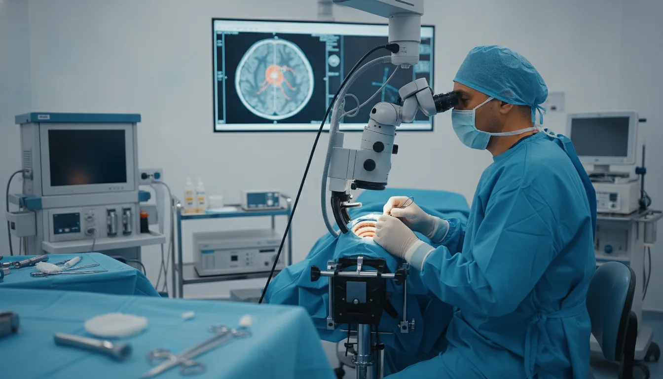
Surgical removal of brain tumors under the operating microscope, guided by neuronavigation for maximum precision.

What is it?

Brain tumor surgery aims to remove the tumor as completely as possible while preserving neurological functions: movement, language, memory and vision. It is a high-precision surgery that now benefits from major technological advances. The operation is planned using a high-resolution pre-operative MRI, which precisely locates the tumor and its relationship to the functional areas of the brain. In the operating room, the neurosurgeon operates under a microscope, guided by neuronavigation — a true surgical GPS — and, when useful, by neurophysiological monitoring, which continuously tracks nerve function throughout the procedure. Every tumor removed is analyzed under the microscope to determine its exact nature. This analysis guides the next steps of care, which are always discussed in a multidisciplinary meeting, in coordination with the other specialists involved. At Clinique Pasteur Tunis, brain tumor surgery relies on a state-of-the-art technical platform — neurosurgical microscope, neuronavigation, intraoperative monitoring, high-resolution MRI — and on a dedicated multipurpose intensive care unit that ensures a safe post-operative course.
Brain tumor surgery

When is it indicated?

Brain tumor detected on imaging and accessible to surgery

When a tumor is visible on MRI and its location allows it to be reached safely, surgery is the first-line treatment to remove it.

Tumor causing headaches, epileptic seizures or neurological deficits

When a tumor causes symptoms that disrupt daily life, its removal aims to make them regress and to protect brain function.

Need for a precise analysis of the lesion (biopsy or resection)

Knowing the exact nature of a lesion is essential for choosing the right treatment. A tissue sample or complete removal allows this analysis under the microscope.

Tumor compressing neighboring structures

A tumor pressing on nearby areas of the brain can impair their function. Removing it relieves this compression and prevents symptoms from worsening.

Preparation

1High-resolution brain MRI for surgical planning
2Complete pre-operative work-up and anesthesia consultation
3Adjustment of ongoing treatments if needed (anticoagulants, antiepileptics)
4Fasting for 6 hours before the operation
5Detailed information for the patient and their family about the procedure and recovery

Procedure

1

General anesthesia with appropriate neurological monitoring

2

Precise positioning and calibration of the neuronavigation system

3

Targeted opening of the skull (craniotomy) over the tumor

4

Removal of the tumor under the operating microscope, guided by neuronavigation

5

Neurophysiological monitoring of nerve function when necessary

6

Careful layer-by-layer closure

7

Post-operative monitoring in intensive care or in a step-down unit

Duration

3 to 6 hours depending on the location

Aftercare & Recovery

A follow-up CT scan or MRI is performed after the operation. Recovery is gradual and may be supported by tailored rehabilitation. The results of the tumor analysis are explained during a consultation and determine the next steps of the care pathway, coordinated by the team.

Risks and side effects

Temporary fatigue and headaches after the operation
Neurological deficit, the risk of which is reduced by neuronavigation and monitoring
Post-operative epileptic seizures, prevented by appropriate treatment
Rare complications inherent to any cranial surgery, managed through intensive care monitoring

These risks remain rare. Your surgeon will inform you in detail before the procedure.

Request a Quote

Contact us to schedule a consultation or receive a personalized quote.

Request a Quote +216 36 402 000

At a glance

Duration: 3 to 6 hours depending on the location
Preparation: Required
Dedicated specialist team