
Transesophageal echocardiography (TEE)
Centres/Cardiac Testing/Transesophageal echo

Transesophageal echocardiography (TEE)

Ultrasound examination of the heart performed by inserting a miniature probe into the esophagus, providing very high-resolution images of the posterior cardiac structures and valves.

What is it?

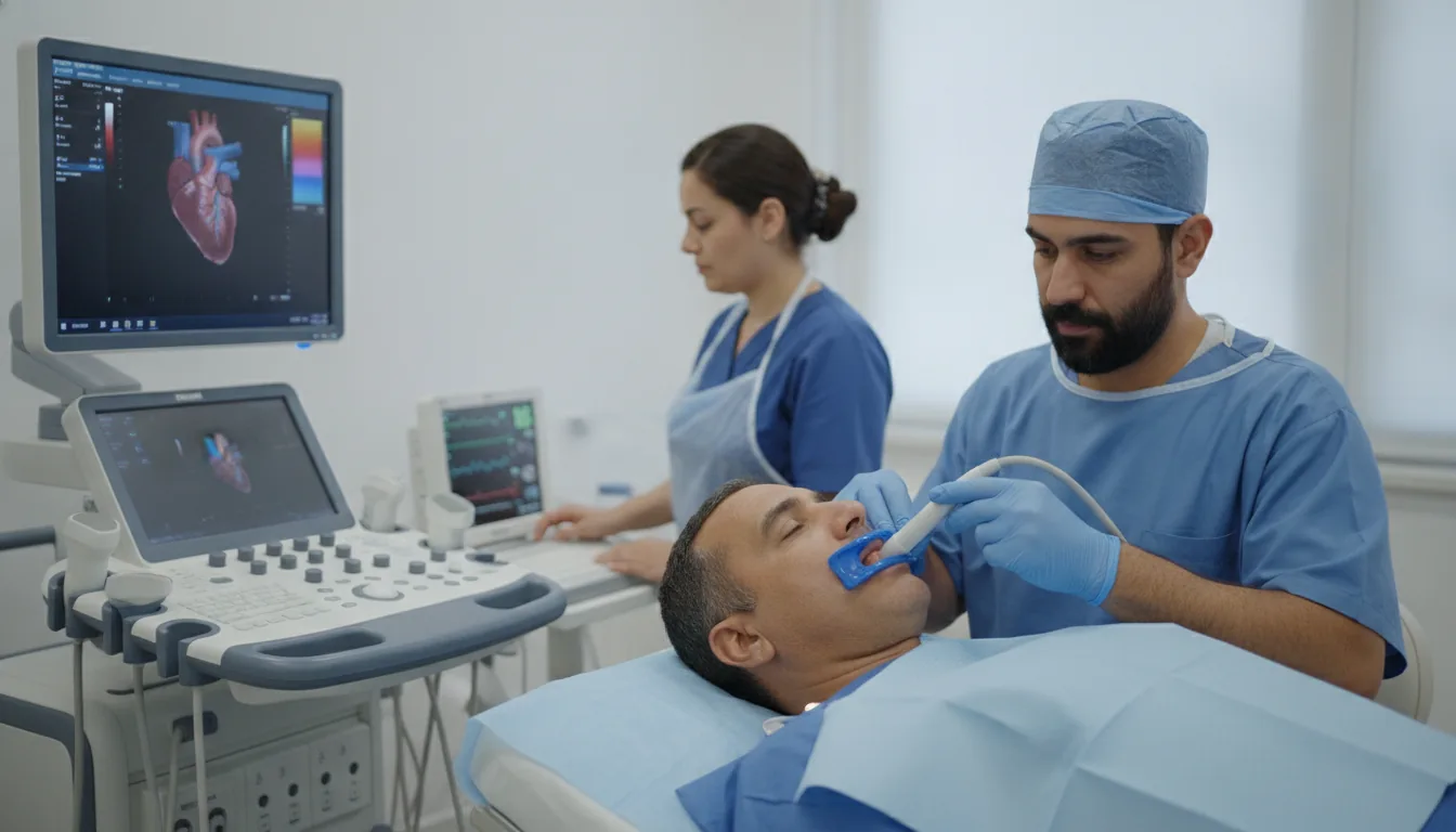
Transesophageal echocardiography is a cardiac imaging examination performed by inserting a miniature ultrasound probe into the esophagus, which lies just behind the heart. This proximity between the probe and the heart produces images of markedly higher resolution than transthoracic echocardiography, particularly for the posterior structures of the heart. TEE is a semi-invasive examination that usually requires local anesthesia of the throat and sometimes light sedation. It is performed when standard transthoracic echocardiography does not provide sufficiently precise images or when certain cardiac structures need to be visualized at maximum resolution. This examination is particularly effective for the detailed assessment of the heart valves (especially the mitral valve), the search for thrombus (clots) in the atria (particularly in the left atrial appendage), the diagnosis of infective endocarditis (valve vegetations), the detection of aortic dissection and the evaluation of prosthetic valves. At the Clinique Pasteur in Tunis, TEE is performed under optimal safety conditions by an experienced echocardiographer, with continuous monitoring of vital signs. Patient comfort is a priority, and light sedation is offered to minimize discomfort during the procedure.
Transesophageal echocardiography (TEE)

When is it indicated?

Search for intra-atrial thrombus before cardioversion

Before restoring a regular heart rhythm with an electric shock, this examination makes sure no clot has formed inside the heart, so the procedure can be carried out safely.

Assessment of infective endocarditis

When an infection of the heart valves is suspected, the probe placed in the esophagus, very close to the heart, provides highly detailed images of any lesions.

Precise assessment of the mitral valve before surgery

Before mitral valve surgery, this examination gives the surgeon a detailed map of the valve to plan the operation as precisely as possible.

Evaluation of prosthetic valves when dysfunction is suspected

If an artificial valve appears to be working less well, ultrasound via the esophagus allows it to be examined very closely and its condition checked.

Search for patent foramen ovale (PFO)

This small passage between the two atria, inherited from fetal life, sometimes remains open; the examination detects it with great accuracy.

Assessment after stroke in young patients

After a stroke in a young person, the examination looks in the heart for a possible cause, such as a clot or a small abnormal communication.

Intraoperative guidance during cardiac surgery

During certain heart operations, this ultrasound guides the surgeon in real time and immediately checks the quality of the result.

Suspected aortic dissection

In an emergency where a tear in the wall of the aorta is suspected, this examination quickly visualizes the artery to confirm or rule out the diagnosis.

Preparation

1Fast strictly for at least 6 hours before the examination
2Report any known allergy, particularly to local anesthetics
3Report any ongoing anticoagulant treatment
4Report any known esophageal condition (esophageal varices, stricture, hiatal hernia)
5Remove removable dentures before the examination
6Arrange for someone to accompany you if sedation is given
7Do not drive for 2 hours after the examination if sedation was given

Procedure

1

An intravenous line is placed for possible administration of sedation

2

The throat is numbed with a local anesthetic spray

3

The patient is positioned lying on their left side

4

A mouthguard is placed between the teeth to protect the probe

5

The ultrasound probe is gently inserted into the esophagus under guidance

6

The cardiologist obtains the various ultrasound views by moving and orienting the probe

7

Color and pulsed Doppler examination is performed

8

The probe is gently withdrawn at the end of the examination

9

The patient remains under observation for 30 minutes to 1 hour after the procedure

Duration

15 to 30 minutes for the examination; allow 1h30 in total including preparation and observation

Results

Results are communicated by the cardiologist within hours of the examination. A detailed report is written and sent to the prescribing physician.

Risks and side effects

Discomfort when the probe is inserted into the throat
Temporary sore throat after the examination (common but harmless)
Superficial esophageal injury (rare)
Esophageal perforation (exceptional, less than 0.01%)
Allergic reaction to the local anesthetic (rare)
Temporary heart rhythm disturbance (rare)

These risks remain rare. Your physician will inform you in detail before the examination.

Book an Appointment

Contact us to schedule your examination or request a quote.

Request a Quote +216 36 402 000

At a glance

Duration: 15 to 30 minutes for the examination; allow 1h30 in total including preparation and observation
Preparation: Required
Book an appointment