
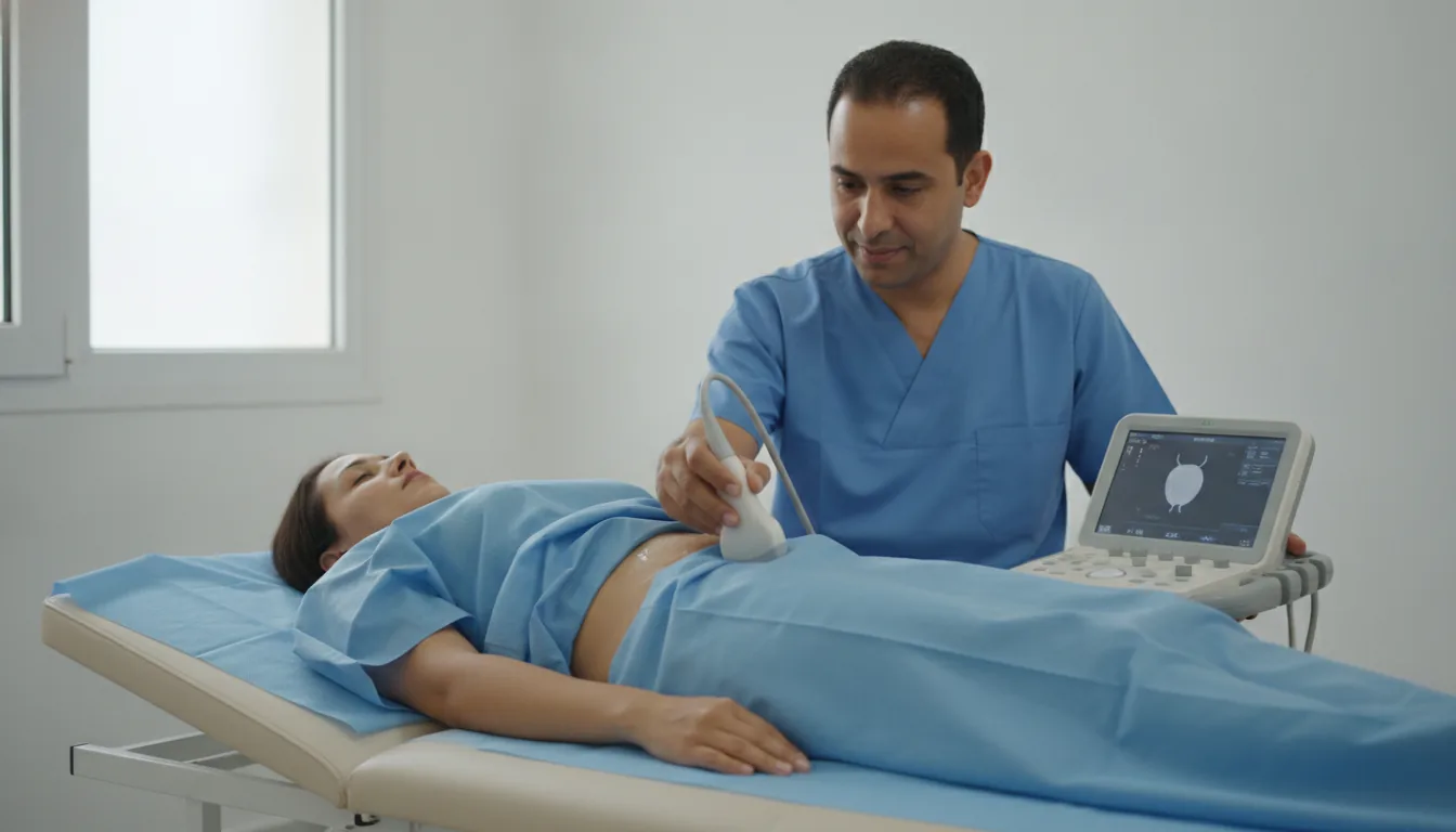
Ultrasound measurement of post-void residual
Centres/Urodynamic Testing/Post-void residual

Ultrasound measurement of post-void residual

Non-invasive assessment by suprapubic ultrasound of the volume of urine remaining in the bladder after voiding.

What is it?

The measurement of the post-void residual (PVR) by ultrasound is a simple, quick and non-invasive test that quantifies the volume of urine remaining in the bladder immediately after voiding. It uses a portable ultrasound scanner or a dedicated device (bladder scanner) placed on the abdomen above the pubic bone. The post-void residual is a fundamental parameter in urology and neuro-urology. A significant residual (generally above 100 ml) may indicate bladder outlet obstruction, detrusor underactivity or a disorder of bladder-sphincter coordination. Its measurement is systematically combined with uroflowmetry. Suprapubic ultrasound not only measures the residual volume but also assesses bladder morphology (wall thickness, presence of diverticula, stones) and detects possible associated kidney abnormalities (pyelocaliceal dilatation). This dual anatomical and functional information makes it an essential first-line test. The measurement is generally performed using the ellipsoid volume formula (length × width × height × 0.52) or automatically by the bladder scanner. Its reliability is excellent and reproducible, making it an ideal monitoring tool for patients under regular urological surveillance.
Ultrasound measurement of post-void residual

When is it indicated?

Systematic complement to uroflowmetry

It accompanies the urinary flow test to check that the bladder empties well after voiding.

Follow-up of patients treated for voiding disorders

It monitors the effectiveness of a treatment by measuring what remains in the bladder after urinating.

Assessment of chronic urinary retention

It helps evaluate a bladder that persistently fails to empty completely.

Monitoring of neurogenic bladders

It is useful for monitoring bladder emptying in people with a neurological disease.

Post-operative follow-up after prostate or pelvic surgery

It verifies that the bladder empties properly after an operation.

Assessment of recurrent urinary tract infections

It helps look for residual urine that could promote repeated urinary tract infections.

Evaluation of bladder emptying disorders

It is offered when the bladder has trouble emptying, to measure the extent of the problem simply.

Preparation

1No specific preparation
2Do not delay voiding before the test: urinate normally
3The measurement is performed immediately after voiding (within 5 minutes)
4No dietary restrictions

Procedure

1

The patient urinates normally (in the toilet or into a flowmeter)

2

Installation lying on the back immediately after voiding

3

Application of ultrasound gel on the abdomen above the pubic bone

4

Measurement of the residual volume by suprapubic ultrasound or bladder scanner

5

Measurements in the three planes of space if a standard ultrasound scanner is used

6

Immediate result displayed on the screen

Duration

5 minutes

Results

The residual volume is expressed in millilitres. A residual below 50 ml is considered normal. Between 50 and 100 ml, it should be monitored. Above 100 ml, it is significant and requires further investigation. The result is immediate.

Risks and side effects

No risk: a completely non-invasive and painless test
No exposure to X-rays (uses ultrasound)
Result may be slightly imprecise in the case of significant obesity

These risks remain rare. Your physician will inform you in detail before the examination.

Book an Appointment

Contact us to schedule your examination or request a quote.

Request a Quote +216 36 402 000

At a glance

Duration: 5 minutes
Preparation: Required
Book an appointment